5CYQ

HIV-1 reverse transcriptase complexed with 4-bromopyrazole


Experimental Data Snapshot

  • Method: X-RAY DIFFRACTION
  • Resolution: 2.15 Å
  • R-Value Free: 0.224 
  • R-Value Work: 0.203 
  • R-Value Observed: 0.204 

wwPDB Validation   3D Report Full Report


Ligand Structure Quality Assessment 


This is version 1.2 of the entry. See complete history


Literature

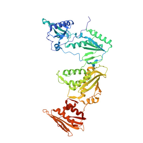
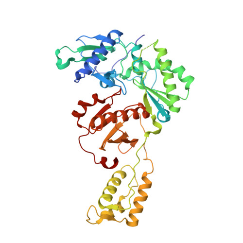
Rapid experimental SAD phasing and hot-spot identification with halogenated fragments.

Bauman, J.D.Harrison, J.J.Arnold, E.

(2016) IUCrJ 3: 51-60

  • DOI: https://doi.org/10.1107/S2052252515021259
  • Primary Citation of Related Structures:  
    5CXR, 5CYQ

  • PubMed Abstract: 

    Through X-ray crystallographic fragment screening, 4-bromopyrazole was discovered to be a 'magic bullet' that is capable of binding at many of the ligand 'hot spots' found in HIV-1 reverse transcriptase (RT). The binding locations can be in pockets that are 'hidden' in the unliganded crystal form, allowing rapid identification of these sites for in silico screening. In addition to hot-spot identification, this ubiquitous yet specific binding provides an avenue for X-ray crystallographic phase determination, which can be a significant bottleneck in the determination of the structures of novel proteins. The anomalous signal from 4-bromopyrazole or 4-iodopyrazole was sufficient to determine the structures of three proteins (HIV-1 RT, influenza A endonuclease and proteinase K) by single-wavelength anomalous dispersion (SAD) from single crystals. Both compounds are inexpensive, readily available, safe and very soluble in DMSO or water, allowing efficient soaking into crystals.


  • Organizational Affiliation

    Center for Advanced Biotechnology and Medicine, Department of Chemistry and Chemical Biology, Rutgers University , 679 Hoes Lane, Piscataway, NJ 08854, USA.


Macromolecules
Find similar proteins by:  (by identity cutoff)  |  3D Structure
Entity ID: 1
MoleculeChains Sequence LengthOrganismDetailsImage
HIV-1 reverse transcriptase, p66 subunit557Human immunodeficiency virus type 1 BH10Mutation(s): 3 
EC: 2.7.7.49
UniProt
Find proteins for P03366 (Human immunodeficiency virus type 1 group M subtype B (isolate BH10))
Explore P03366 
Go to UniProtKB:  P03366
Entity Groups  
Sequence Clusters30% Identity50% Identity70% Identity90% Identity95% Identity100% Identity
UniProt GroupP03366
Sequence Annotations
Expand
  • Reference Sequence
Find similar proteins by:  (by identity cutoff)  |  3D Structure
Entity ID: 2
MoleculeChains Sequence LengthOrganismDetailsImage
HIV-1 reverse transcriptase, p51 subunit428Human immunodeficiency virus type 1 BH10Mutation(s): 1 
EC: 2.7.7.49
UniProt
Find proteins for P03366 (Human immunodeficiency virus type 1 group M subtype B (isolate BH10))
Explore P03366 
Go to UniProtKB:  P03366
Entity Groups  
Sequence Clusters30% Identity50% Identity70% Identity90% Identity95% Identity100% Identity
UniProt GroupP03366
Sequence Annotations
Expand
  • Reference Sequence
Small Molecules
Ligands 4 Unique
IDChains Name / Formula / InChI Key2D Diagram3D Interactions
T27
Query on T27

Download Ideal Coordinates CCD File 
C [auth A]4-{[4-({4-[(E)-2-cyanoethenyl]-2,6-dimethylphenyl}amino)pyrimidin-2-yl]amino}benzonitrile
C22 H18 N6
YIBOMRUWOWDFLG-ONEGZZNKSA-N
BYZ
Query on BYZ

Download Ideal Coordinates CCD File 
D [auth A]
E [auth A]
F [auth A]
G [auth A]
H [auth A]
D [auth A],
E [auth A],
F [auth A],
G [auth A],
H [auth A],
I [auth A],
J [auth A],
K [auth A],
S [auth B],
T [auth B],
U [auth B],
V [auth B],
W [auth B],
X [auth B],
Y [auth B]
4-bromo-1H-pyrazole
C3 H3 Br N2
WVGCPEDBFHEHEZ-UHFFFAOYSA-N
BR
Query on BR

Download Ideal Coordinates CCD File 
P [auth A],
Q [auth A],
R [auth A]
BROMIDE ION
Br
CPELXLSAUQHCOX-UHFFFAOYSA-M
DMS
Query on DMS

Download Ideal Coordinates CCD File 
AA [auth B]
BA [auth B]
CA [auth B]
DA [auth B]
EA [auth B]
AA [auth B],
BA [auth B],
CA [auth B],
DA [auth B],
EA [auth B],
L [auth A],
M [auth A],
N [auth A],
O [auth A],
Z [auth B]
DIMETHYL SULFOXIDE
C2 H6 O S
IAZDPXIOMUYVGZ-UHFFFAOYSA-N
Binding Affinity Annotations 
IDSourceBinding Affinity
T27 BindingDB:  5CYQ IC50: min: 38, max: 1.00e+4 (nM) from 3 assay(s)
Experimental Data & Validation

Experimental Data

  • Method: X-RAY DIFFRACTION
  • Resolution: 2.15 Å
  • R-Value Free: 0.224 
  • R-Value Work: 0.203 
  • R-Value Observed: 0.204 
  • Space Group: C 1 2 1
Unit Cell:
Length ( Å )Angle ( ˚ )
a = 161.887α = 90
b = 73.167β = 100.56
c = 108.894γ = 90
Software Package:
Software NamePurpose
PHENIXrefinement
DENZOdata reduction
SCALEPACKdata scaling
PHENIXphasing

Structure Validation

View Full Validation Report


Ligand Structure Quality Assessment 


Entry History 

Deposition Data

Revision History  (Full details and data files)

  • Version 1.0: 2015-12-30
    Type: Initial release
  • Version 1.1: 2018-04-18
    Changes: Data collection, Database references, Derived calculations
  • Version 1.2: 2023-09-27
    Changes: Data collection, Database references, Refinement description