5CGO

Structure of quasiracemic Ala-Magainin 2 with a beta amino acid substitution at position 13


Experimental Data Snapshot

  • Method: X-RAY DIFFRACTION
  • Resolution: 1.50 Å
  • R-Value Free: 0.239 
  • R-Value Work: 0.172 
  • R-Value Observed: 0.175 

wwPDB Validation   3D Report Full Report


This is version 1.6 of the entry. See complete history


Literature

Quasiracemate Crystal Structures of Magainin 2 Derivatives Support the Functional Significance of the Phenylalanine Zipper Motif.

Hayouka, Z.Thomas, N.C.Mortenson, D.E.Satyshur, K.A.Weisblum, B.Forest, K.T.Gellman, S.H.

(2015) J Am Chem Soc 137: 11884-11887

  • DOI: https://doi.org/10.1021/jacs.5b07206
  • Primary Citation of Related Structures:  
    5CGN, 5CGO

  • PubMed Abstract: 

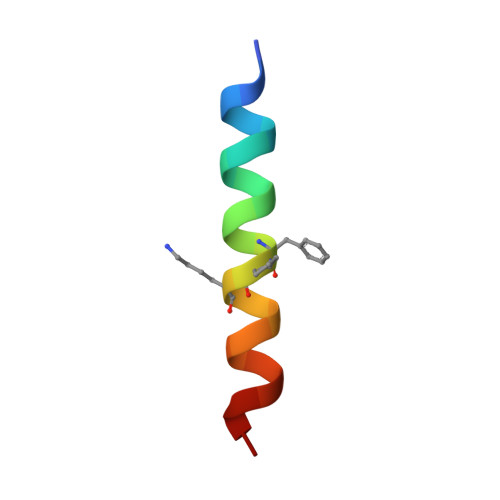
    Quasiracemic crystallography has been used to explore the significance of homochiral and heterochiral associations in a set of host-defense peptide derivatives. The previously reported racemic crystal structure of a magainin 2 derivative displayed a homochiral antiparallel dimer association featuring a "phenylalanine zipper" notable for the dual roles of phenylalanines in mediating dimerization and formation of an exposed hydrophobic swath. This motif is seen as well in two new quasiracemate crystals that contain the d form of the magainin 2 derivative along with an l-peptide in which one Ala has been replaced by a β-amino acid residue. This structural trend supports the hypothesis that the Phe zipper motif has functional significance.


  • Organizational Affiliation

    Department of Chemistry, ‡Department of Medicine, and §Department of Bacteriology, University of Wisconsin-Madison , Madison, Wisconsin 53706, United States.


Macromolecules

Find similar proteins by:  Sequence   |   3D Structure  

Entity ID: 1
MoleculeChains Sequence LengthOrganismDetailsImage
ACPC-13 derivative of Ala-Magainin 2
A, B
23synthetic constructMutation(s): 0 
Membrane Entity: Yes 
UniProt
Find proteins for P11006 (Xenopus laevis)
Explore P11006 
Go to UniProtKB:  P11006
Entity Groups  
Sequence Clusters30% Identity50% Identity70% Identity90% Identity95% Identity100% Identity
UniProt GroupP11006
Sequence Annotations
Expand
  • Reference Sequence

Find similar proteins by:  Sequence   |   3D Structure  

Entity ID: 2
MoleculeChains Sequence LengthOrganismDetailsImage
D-Ala-Magainin 2
C, D
23synthetic constructMutation(s): 0 
Sequence Annotations
Expand
  • Reference Sequence
Experimental Data & Validation

Experimental Data

  • Method: X-RAY DIFFRACTION
  • Resolution: 1.50 Å
  • R-Value Free: 0.239 
  • R-Value Work: 0.172 
  • R-Value Observed: 0.175 
  • Space Group: P 1
Unit Cell:
Length ( Å )Angle ( ˚ )
a = 24.85α = 109.76
b = 27β = 102.35
c = 29.96γ = 90.23
Software Package:
Software NamePurpose
REFMACrefinement
XDSdata reduction
XSCALEdata scaling
PHASERphasing

Structure Validation

View Full Validation Report


Entry History & Funding Information

Deposition Data


Funding OrganizationLocationGrant Number
National Institutes of Health/National Institute of General Medical Sciences (NIH/NIGMS)United StatesR01 GM061238

Revision History  (Full details and data files)

  • Version 1.0: 2015-09-23
    Type: Initial release
  • Version 1.1: 2015-09-30
    Changes: Database references
  • Version 1.2: 2015-10-07
    Changes: Database references
  • Version 1.3: 2017-09-20
    Changes: Author supporting evidence, Derived calculations
  • Version 1.4: 2019-12-25
    Changes: Author supporting evidence
  • Version 1.5: 2023-09-27
    Changes: Data collection, Database references, Refinement description
  • Version 1.6: 2023-11-15
    Changes: Data collection, Derived calculations