5BS1

Crystal structure of RbcX-IIa from Chlamydomonas reinhardtii


Experimental Data Snapshot

  • Method: X-RAY DIFFRACTION
  • Resolution: 1.60 Å
  • R-Value Free: 0.206 
  • R-Value Work: 0.177 
  • R-Value Observed: 0.179 

wwPDB Validation   3D Report Full Report


This is version 1.1 of the entry. See complete history


Literature

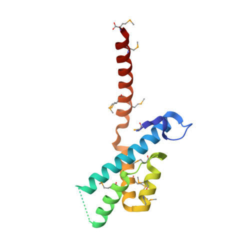
Structural Analysis of the Rubisco-Assembly Chaperone RbcX-II from Chlamydomonas reinhardtii.

Bracher, A.Hauser, T.Liu, C.Hartl, F.U.Hayer-Hartl, M.

(2015) PLoS One 10: e0135448-e0135448

  • DOI: https://doi.org/10.1371/journal.pone.0135448
  • Primary Citation of Related Structures:  
    5BS1, 5BS2

  • PubMed Abstract: 

    The most prevalent form of the Rubisco enzyme is a complex of eight catalytic large subunits (RbcL) and eight regulatory small subunits (RbcS). Rubisco biogenesis depends on the assistance by specific molecular chaperones. The assembly chaperone RbcX stabilizes the RbcL subunits after folding by chaperonin and mediates their assembly to the RbcL8 core complex, from which RbcX is displaced by RbcS to form active holoenzyme. Two isoforms of RbcX are found in eukaryotes, RbcX-I, which is more closely related to cyanobacterial RbcX, and the more distant RbcX-II. The green algae Chlamydomonas reinhardtii contains only RbcX-II isoforms, CrRbcX-IIa and CrRbcX-IIb. Here we solved the crystal structure of CrRbcX-IIa and show that it forms an arc-shaped dimer with a central hydrophobic cleft for binding the C-terminal sequence of RbcL. Like other RbcX proteins, CrRbcX-IIa supports the assembly of cyanobacterial Rubisco in vitro, albeit with reduced activity relative to cyanobacterial RbcX-I. Structural analysis of a fusion protein of CrRbcX-IIa and the C-terminal peptide of RbcL suggests that the peptide binding mode of RbcX-II may differ from that of cyanobacterial RbcX. RbcX homologs appear to have adapted to their cognate Rubisco clients as a result of co-evolution.


  • Organizational Affiliation

    Department of Cellular Biochemistry, Max-Planck-Institute of Biochemistry, Martinsried, Germany.


Macromolecules
Find similar proteins by:  (by identity cutoff)  |  3D Structure
Entity ID: 1
MoleculeChains Sequence LengthOrganismDetailsImage
CrRbcX-IIa
A, B, C, D
123Chlamydomonas reinhardtiiMutation(s): 0 
Gene Names: CGL41CHLREDRAFT_162607
Entity Groups  
Sequence Clusters30% Identity50% Identity70% Identity90% Identity95% Identity100% Identity
Sequence Annotations
Expand
  • Reference Sequence
Small Molecules
Ligands 1 Unique
IDChains Name / Formula / InChI Key2D Diagram3D Interactions
MG
Query on MG

Download Ideal Coordinates CCD File 
E [auth B]MAGNESIUM ION
Mg
JLVVSXFLKOJNIY-UHFFFAOYSA-N
Modified Residues  1 Unique
IDChains TypeFormula2D DiagramParent
MSE
Query on MSE
A, B, C, D
L-PEPTIDE LINKINGC5 H11 N O2 SeMET
Experimental Data & Validation

Experimental Data

  • Method: X-RAY DIFFRACTION
  • Resolution: 1.60 Å
  • R-Value Free: 0.206 
  • R-Value Work: 0.177 
  • R-Value Observed: 0.179 
  • Space Group: P 1
Unit Cell:
Length ( Å )Angle ( ˚ )
a = 36.133α = 76.49
b = 52.989β = 81.1
c = 61.561γ = 70.1
Software Package:
Software NamePurpose
SCALAdata scaling
SHELXphasing
REFMACrefinement
PDB_EXTRACTdata extraction
XDSdata reduction

Structure Validation

View Full Validation Report


Entry History 

Deposition Data

Revision History  (Full details and data files)

  • Version 1.0: 2015-08-05
    Type: Initial release
  • Version 1.1: 2015-09-09
    Changes: Database references