5BR1

CRYSTAL STRUCTURE OF AN ABC TRANSPORTER SOLUTE BINDING PROTEIN (IPR025997) FROM AGROBACTERIUM VITIS S4 (Avi_5305, TARGET EFI-511224) WITH BOUND ALPHA-D-GALACTOSAMINE


Experimental Data Snapshot

  • Method: X-RAY DIFFRACTION
  • Resolution: 1.85 Å
  • R-Value Free: 0.283 
  • R-Value Work: 0.183 
  • R-Value Observed: 0.188 

Starting Model: experimental
View more details

wwPDB Validation   3D Report Full Report


Ligand Structure Quality Assessment 


This is version 2.2 of the entry. See complete history


Literature

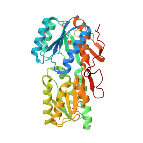
Structure of an ABC transporter solute-binding protein specific for the amino sugars glucosamine and galactosamine.

Yadava, U.Vetting, M.W.Al Obaidi, N.Carter, M.S.Gerlt, J.A.Almo, S.C.

(2016) Acta Crystallogr F Struct Biol Commun 72: 467-472

  • DOI: https://doi.org/10.1107/S2053230X16007500
  • Primary Citation of Related Structures:  
    4Y9T, 5BR1

  • PubMed Abstract: 

    The uptake of exogenous solutes by prokaryotes is mediated by transport systems embedded in the plasma membrane. In many cases, a solute-binding protein (SBP) is utilized to bind ligands with high affinity and deliver them to the membrane-bound components responsible for translocation into the cytoplasm. In the present study, Avi_5305, an Agrobacterium vitis SBP belonging to Pfam13407, was screened by differential scanning fluorimetry (DSF) and found to be stabilized by D-glucosamine and D-galactosamine. Avi_5305 is the first protein from Pfam13407 shown to be specific for amino sugars, and co-crystallization resulted in structures of Avi_5305 bound to D-glucosamine and D-galactosamine. Typical of Pfam13407, Avi_5305 consists of two α/β domains linked through a hinge region, with the ligand-binding site located in a cleft between the two domains. Comparisons with Escherichia coli ribose-binding protein suggest that a cation-π interaction with Tyr168 provides the specificity for D-glucosamine/D-galactosamine over D-glucose/D-galactose.


  • Organizational Affiliation

    Department of Physics, DDU Gorakhpur University, Gorakhpur 273 009, India.


Macromolecules
Find similar proteins by:  (by identity cutoff)  |  3D Structure
Entity ID: 1
MoleculeChains Sequence LengthOrganismDetailsImage
ABC transporter, binding protein346Allorhizobium ampelinum S4Mutation(s): 0 
Gene Names: Avi_5305
UniProt
Find proteins for B9K0Q5 (Allorhizobium ampelinum (strain ATCC BAA-846 / DSM 112012 / S4))
Explore B9K0Q5 
Go to UniProtKB:  B9K0Q5
Entity Groups  
Sequence Clusters30% Identity50% Identity70% Identity90% Identity95% Identity100% Identity
UniProt GroupB9K0Q5
Sequence Annotations
Expand
  • Reference Sequence
Small Molecules
Ligands 1 Unique
IDChains Name / Formula / InChI Key2D Diagram3D Interactions
X6X
Query on X6X

Download Ideal Coordinates CCD File 
B [auth A]2-amino-2-deoxy-alpha-D-galactopyranose
C6 H13 N O5
MSWZFWKMSRAUBD-DVKNGEFBSA-N
Modified Residues  1 Unique
IDChains TypeFormula2D DiagramParent
MSE
Query on MSE
A
L-PEPTIDE LINKINGC5 H11 N O2 SeMET
Experimental Data & Validation

Experimental Data

  • Method: X-RAY DIFFRACTION
  • Resolution: 1.85 Å
  • R-Value Free: 0.283 
  • R-Value Work: 0.183 
  • R-Value Observed: 0.188 
  • Space Group: P 21 21 21
Unit Cell:
Length ( Å )Angle ( ˚ )
a = 36.781α = 90
b = 63.701β = 90
c = 119.575γ = 90
Software Package:
Software NamePurpose
HKL-2000data collection
HKL-2000data scaling
PHENIXrefinement
PDB_EXTRACTdata extraction
PHENIXphasing
HKL-3000data reduction

Structure Validation

View Full Validation Report


Ligand Structure Quality Assessment 


Entry History 

Revision History  (Full details and data files)

  • Version 1.0: 2015-06-10
    Type: Initial release
  • Version 1.1: 2016-06-22
    Changes: Database references
  • Version 2.0: 2020-07-29
    Type: Remediation
    Reason: Carbohydrate remediation
    Changes: Atomic model, Data collection, Derived calculations
  • Version 2.1: 2023-09-27
    Changes: Data collection, Database references, Refinement description, Structure summary
  • Version 2.2: 2023-11-15
    Changes: Data collection