All Ca(2+)-binding loops of light-sensitive ctenophore photoprotein berovin bind magnesium ions: The spatial structure of Mg(2+)-loaded apo-berovin.
Burakova, L.P., Natashin, P.V., Malikova, N.P., Niu, F., Pu, M., Vysotski, E.S., Liu, Z.J.(2016) J Photochem Photobiol B 154: 57-66
- PubMed: 26690016
- DOI: https://doi.org/10.1016/j.jphotobiol.2015.11.012
- Primary Citation of Related Structures:
5BPJ - PubMed Abstract:
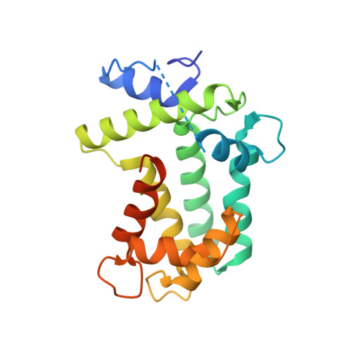
Light-sensitive photoprotein berovin accounts for a bright bioluminescence of ctenophore Beroe abyssicola. Berovin is functionally identical to the well-studied Ca(2+)-regulated photoproteins of jellyfish, however in contrast to those it is extremely sensitive to the visible light. Berovin contains three EF-hand Ca(2+)-binding sites and consequently belongs to a large family of the EF-hand Ca(2+)-binding proteins. Here we report the spatial structure of apo-berovin with bound Mg(2+) determined at 1.75Å. The magnesium ion is found in each functional EF-hand loop of a photoprotein and coordinated by oxygen atoms donated by the side-chain groups of aspartate, carbonyl groups of the peptide backbone, or hydroxyl group of serine with characteristic oxygen-Mg(2+) distances. As oxygen supplied by the side-chain of the twelfth residue of all Ca(2+)-binding loops participates in the magnesium ion coordination, it was suggested that Ca(2+)-binding loops of berovin belong to the mixed Ca(2+)/Mg(2+) rather than Ca(2+)-specific type. In addition, we report an effect of physiological concentration of Mg(2+) on bioluminescence of berovin (sensitivity to Ca(2+), rapid-mixed kinetics, light-sensitivity, thermostability, and apo-berovin conversion into active protein). The different impact of physiological concentration of Mg(2+) on berovin bioluminescence as compared to hydromedusan photoproteins was attributed to different affinities of the Ca(2+)-binding sites of these photoproteins to Mg(2+).
Organizational Affiliation:
Institute of Molecular and Clinical Medicine, Kunming Medical University, Kunming 650500, China; Photobiology Laboratory, Institute of Biophysics, Russian Academy of Sciences, Siberian Branch, Akademgorodok 50, Bldg. 50, Krasnoyarsk 660036, Russia; National Laboratory of Biomacromolecules, Institute of Biophysics, Chinese Academy of Sciences, 15 Datun Road, Beijing 100101, China.