5BOI

Bacillus megaterium YpeB C-terminal domain


Experimental Data Snapshot

  • Method: X-RAY DIFFRACTION
  • Resolution: 1.80 Å
  • R-Value Free: 0.227 
  • R-Value Work: 0.195 
  • R-Value Observed: 0.198 

wwPDB Validation   3D Report Full Report


This is version 1.1 of the entry. See complete history


Literature

Crystal structure of the PepSY-containing domain of the YpeB protein involved in germination of bacillus spores.

Ustok, F.I.Chirgadze, D.Y.Christie, G.

(2015) Proteins 83: 1914-1921

  • DOI: https://doi.org/10.1002/prot.24868
  • Primary Citation of Related Structures:  
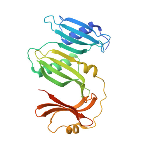
    5BOI

  • PubMed Abstract: 

    The crystal structure of the C-terminal domain of the Bacillus megaterium YpeB protein has been solved by X-ray crystallography to 1.80-Å resolution. The full-length protein is essential in stabilising the SleB cortex lytic enzyme in Bacillus spores, and may have a role in regulating SleB activity during spore germination. The YpeB-C crystal structure comprises three tandemly repeated PepSY domains, which are aligned to form an extended laterally compressed molecule. A predominantly positively charged region located in the second PepSY domain may provide a site for protein interactions that are important in stabilising SleB and YpeB within the spore.


  • Organizational Affiliation

    Department of Chemical Engineering and Biotechnology, Institute of Biotechnology, University of Cambridge, Cambridge, United Kingdom.


Macromolecules
Find similar proteins by:  (by identity cutoff)  |  3D Structure
Entity ID: 1
MoleculeChains Sequence LengthOrganismDetailsImage
Germination protein YpeB246Priestia megaterium QM B1551Mutation(s): 0 
Gene Names: ypeBBMQ_4349
UniProt
Find proteins for D5DRI0 (Priestia megaterium (strain ATCC 12872 / QMB1551))
Explore D5DRI0 
Go to UniProtKB:  D5DRI0
Entity Groups  
Sequence Clusters30% Identity50% Identity70% Identity90% Identity95% Identity100% Identity
UniProt GroupD5DRI0
Sequence Annotations
Expand
  • Reference Sequence
Experimental Data & Validation

Experimental Data

  • Method: X-RAY DIFFRACTION
  • Resolution: 1.80 Å
  • R-Value Free: 0.227 
  • R-Value Work: 0.195 
  • R-Value Observed: 0.198 
  • Space Group: C 2 2 21
Unit Cell:
Length ( Å )Angle ( ˚ )
a = 53.208α = 90
b = 110.104β = 90
c = 85.806γ = 90
Software Package:
Software NamePurpose
PHENIXrefinement
XDSdata reduction
XDSdata scaling
PHASERphasing

Structure Validation

View Full Validation Report


Entry History 

Deposition Data

Revision History  (Full details and data files)

  • Version 1.0: 2015-08-12
    Type: Initial release
  • Version 1.1: 2015-09-16
    Changes: Database references