5B8C

High resolution structure of the human PD-1 in complex with pembrolizumab Fv


Experimental Data Snapshot

  • Method: X-RAY DIFFRACTION
  • Resolution: 2.15 Å
  • R-Value Free: 0.226 
  • R-Value Work: 0.184 
  • R-Value Observed: 0.186 

wwPDB Validation   3D Report Full Report


This is version 1.3 of the entry. See complete history


Literature

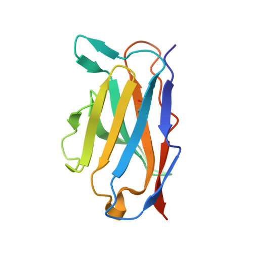
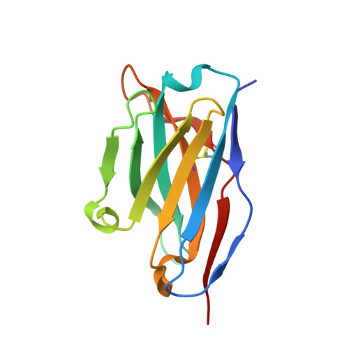
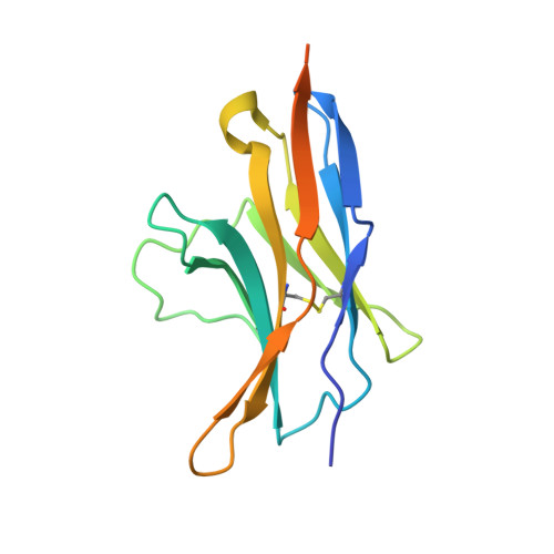
High-resolution crystal structure of the therapeutic antibody pembrolizumab bound to the human PD-1

Horita, S.Nomura, Y.Sato, Y.Shimamura, T.Iwata, S.Nomura, N.

(2016) Sci Rep 6: 35297-35297

  • DOI: https://doi.org/10.1038/srep35297
  • Primary Citation of Related Structures:  
    5B8C

  • PubMed Abstract: 

    Pembrolizumab is an FDA-approved therapeutic antibody that targets the programmed cell death-1 (PD-1) to block the immune checkpoint pathway for the treatment of various types of cancer. It receives remarkable attention due to the high degree of efficacy. Very recently, the crystal structure of the Fab fragment of pembrolizumab (PemFab) in complex with the extracellular domain of human PD-1 (PD-1 ECD ) was reported at a resolution of 2.9 Å. However, this relatively low-resolution structural data fails to provide sufficient information on interfacial water molecules at the binding interface that substantially contribute to affinity and specificity between the therapeutic antibody and target. Here, we present the independently determined crystal structure of the Fv fragment of pembrolizumab (PemFv) in complex with the PD-1 ECD at a resolution of 2.15 Å. This high-resolution structure allows the accurate mapping of the interaction including water-mediated hydrogen bonds and provides, for the first time, a coherent explanation of PD-1 antagonism by pembrolizumab. Our structural data also provides new insights into the rational design of improved anti-PD-1 therapeutics.


  • Organizational Affiliation

    Department of Cell Biology, Graduate School of Medicine, Kyoto University, Yoshida-Konoe-cho, Sakyo-ku, Kyoto 606-8501, Japan.


Macromolecules
Find similar proteins by:  (by identity cutoff)  |  3D Structure
Entity ID: 1
MoleculeChains Sequence LengthOrganismDetailsImage
Pembrolizumab light chain variable region (PemVL)
A, D, G, J
119Homo sapiensMutation(s): 0 
Entity Groups  
Sequence Clusters30% Identity50% Identity70% Identity90% Identity95% Identity100% Identity
Sequence Annotations
Expand
  • Reference Sequence
Find similar proteins by:  (by identity cutoff)  |  3D Structure
Entity ID: 2
MoleculeChains Sequence LengthOrganismDetailsImage
Pembrolizumab heavy chain variable region (PemVH)
B, E, H, K
120Homo sapiensMutation(s): 0 
Entity Groups  
Sequence Clusters30% Identity50% Identity70% Identity90% Identity95% Identity100% Identity
Sequence Annotations
Expand
  • Reference Sequence
Find similar proteins by:  (by identity cutoff)  |  3D Structure
Entity ID: 3
MoleculeChains Sequence LengthOrganismDetailsImage
Programmed cell death protein 1
C, F, I, L
139Homo sapiensMutation(s): 1 
Gene Names: PDCD1PD1
UniProt & NIH Common Fund Data Resources
Find proteins for Q15116 (Homo sapiens)
Explore Q15116 
Go to UniProtKB:  Q15116
PHAROS:  Q15116
GTEx:  ENSG00000188389 
Entity Groups  
Sequence Clusters30% Identity50% Identity70% Identity90% Identity95% Identity100% Identity
UniProt GroupQ15116
Sequence Annotations
Expand
  • Reference Sequence
Experimental Data & Validation

Experimental Data

  • Method: X-RAY DIFFRACTION
  • Resolution: 2.15 Å
  • R-Value Free: 0.226 
  • R-Value Work: 0.184 
  • R-Value Observed: 0.186 
  • Space Group: P 21 21 21
Unit Cell:
Length ( Å )Angle ( ˚ )
a = 143.68α = 90
b = 143.13β = 90
c = 76.55γ = 90
Software Package:
Software NamePurpose
PHENIXrefinement
XDSdata reduction
XDSdata scaling
PHASERphasing

Structure Validation

View Full Validation Report


Entry History & Funding Information

Deposition Data


Funding OrganizationLocationGrant Number
Japan Science and Technology Agency (JST)Japan--
Ministry of Education, Culture, Sports, Science and Technology (MEXT) of JapanJapan--
Japan Society for the Promotion of ScienceJapan15K06968
Japan Society for the Promotion of ScienceJapan15J04343

Revision History  (Full details and data files)

  • Version 1.0: 2016-10-26
    Type: Initial release
  • Version 1.1: 2017-09-27
    Changes: Data collection, Derived calculations
  • Version 1.2: 2017-10-18
    Changes: Author supporting evidence
  • Version 1.3: 2023-11-08
    Changes: Data collection, Database references, Refinement description