SARS-CoV 3CL protease cleaves its C-terminal autoprocessing site by novel subsite cooperativity
Muramatsu, T., Takemoto, C., Kim, Y.T., Wang, H., Nishii, W., Terada, T., Shirouzu, M., Yokoyama, S.(2016) Proc Natl Acad Sci U S A 113: 12997-13002
- PubMed: 27799534
- DOI: https://doi.org/10.1073/pnas.1601327113
- Primary Citation of Related Structures:
2DUC, 5B6O - PubMed Abstract:
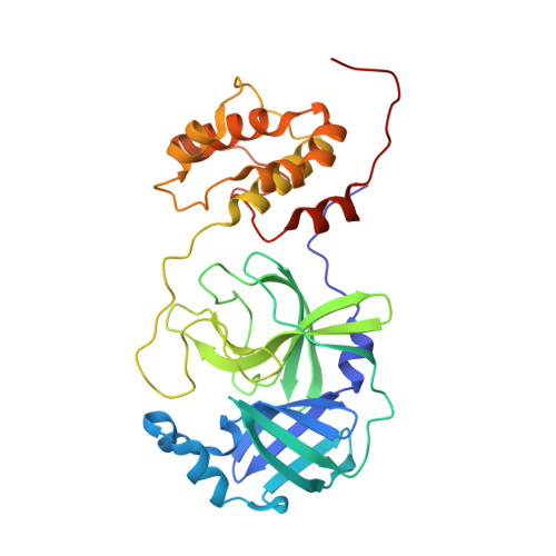
The 3C-like protease (3CL pro ) of severe acute respiratory syndrome coronavirus (SARS-CoV) cleaves 11 sites in the polyproteins, including its own N- and C-terminal autoprocessing sites, by recognizing P4-P1 and P1'. In this study, we determined the crystal structure of 3CL pro with the C-terminal prosequence and the catalytic-site C145A mutation, in which the enzyme binds the C-terminal prosequence of another molecule. Surprisingly, Phe at the P3' position [Phe(P3')] is snugly accommodated in the S3' pocket. Mutations of Phe(P3') impaired the C-terminal autoprocessing, but did not affect N-terminal autoprocessing. This difference was ascribed to the P2 residue, Phe(P2) and Leu(P2), in the C- and N-terminal sites, as follows. The S3' subsite is formed by Phe(P2)-induced conformational changes of 3CL pro and the direct involvement of Phe(P2) itself. In contrast, the N-terminal prosequence with Leu(P2) does not cause such conformational changes for the S3' subsite formation. In fact, the mutation of Phe(P2) to Leu in the C-terminal autoprocessing site abolishes the dependence on Phe(P3'). These mechanisms explain why Phe is required at the P3' position when the P2 position is occupied by Phe rather than Leu, which reveals a type of subsite cooperativity. Moreover, the peptide consisting of P4-P1 with Leu(P2) inhibits protease activity, whereas that with Phe(P2) exhibits a much smaller inhibitory effect, because Phe(P3') is missing. Thus, this subsite cooperativity likely exists to avoid the autoinhibition of the enzyme by its mature C-terminal sequence, and to retain the efficient C-terminal autoprocessing by the use of Phe(P2).
Organizational Affiliation:
RIKEN Systems and Structural Biology Center, Tsurumi, Yokohama 230-0045, Japan; tomonari.muramatsu@riken.jp yokoyama@riken.jp.