5AF2

Crystal structure of the C-terminal 2',5'-phosphodiesterase domain of group A rotavirus protein VP3


Experimental Data Snapshot

  • Method: X-RAY DIFFRACTION
  • Resolution: 1.39 Å
  • R-Value Free: 0.193 
  • R-Value Work: 0.171 
  • R-Value Observed: 0.172 

wwPDB Validation   3D Report Full Report


This is version 1.3 of the entry. See complete history


Literature

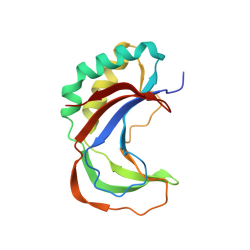
Crystal Structure of the C-Terminal 2',5'-Phosphodiesterase Domain of Group a Rotavirus Protein Vp3.

Brandmann, T.Jinek, M.

(2015) Proteins 83: 997

  • DOI: https://doi.org/10.1002/prot.24794
  • Primary Citation of Related Structures:  
    5AF2

  • PubMed Abstract: 

    In response to viral infections, the mammalian innate immune system induces the production of the second messenger 2'-5' oligoadenylate (2-5A) to activate latent ribonuclease L (RNase L) that restricts viral replication and promotes apoptosis. A subset of rotaviruses and coronaviruses encode 2',5'-phosphodiesterase enzymes that hydrolyze 2-5A, thereby inhibiting RNase L activation. We report the crystal structure of the 2',5'-phosphodiesterase domain of group A rotavirus protein VP3 at 1.39 Å resolution. The structure exhibits a 2H phosphoesterase fold and reveals conserved active site residues, providing insights into the mechanism of 2-5A degradation in viral evasion of host innate immunity.


  • Organizational Affiliation

    Department of Biochemistry, University of Zurich, Zurich, Switzerland.


Macromolecules
Find similar proteins by:  (by identity cutoff)  |  3D Structure
Entity ID: 1
MoleculeChains Sequence LengthOrganismDetailsImage
VP3
A, B, C, D
143Rotavirus AMutation(s): 0 
UniProt
Find proteins for A2T3S5 (Rotavirus A (isolate RVA/Monkey/South Africa/SA11-H96/1958/G3P5B[2]))
Explore A2T3S5 
Go to UniProtKB:  A2T3S5
Entity Groups  
Sequence Clusters30% Identity50% Identity70% Identity90% Identity95% Identity100% Identity
UniProt GroupA2T3S5
Sequence Annotations
Expand
  • Reference Sequence
Experimental Data & Validation

Experimental Data

  • Method: X-RAY DIFFRACTION
  • Resolution: 1.39 Å
  • R-Value Free: 0.193 
  • R-Value Work: 0.171 
  • R-Value Observed: 0.172 
  • Space Group: P 1 21 1
Unit Cell:
Length ( Å )Angle ( ˚ )
a = 61.29α = 90
b = 74.96β = 112.8
c = 63.22γ = 90
Software Package:
Software NamePurpose
PHENIXrefinement
XDSdata reduction
SCALAdata scaling
PHENIXphasing

Structure Validation

View Full Validation Report


Entry History 

Deposition Data

Revision History  (Full details and data files)

  • Version 1.0: 2015-03-18
    Type: Initial release
  • Version 1.1: 2015-03-25
    Changes: Database references
  • Version 1.2: 2015-04-08
    Changes: Atomic model
  • Version 1.3: 2015-04-29
    Changes: Database references