5A9K

Structural basis for DNA strand separation by a hexameric replicative helicase


Experimental Data Snapshot

  • Method: ELECTRON MICROSCOPY
  • Resolution: 19.0 Å
  • Aggregation State: PARTICLE 
  • Reconstruction Method: SINGLE PARTICLE 

wwPDB Validation   3D Report Full Report


This is version 1.2 of the entry. See complete history


Literature

Structural Basis for DNA Strand Separation by a Hexameric Replicative Helicase.

Chaban, Y.Stead, J.A.Ryzhenkova, K.Whelan, F.Lamber, K.Antson, A.Sanders, C.M.Orlova, E.V.

(2015) Nucleic Acids Res 43: 8551

  • DOI: https://doi.org/10.1093/nar/gkv778
  • Primary Citation of Related Structures:  
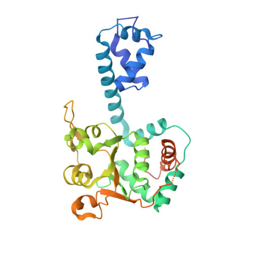
    5A9K

  • PubMed Abstract: 

    Hexameric helicases are processive DNA unwinding machines but how they engage with a replication fork during unwinding is unknown. Using electron microscopy and single particle analysis we determined structures of the intact hexameric helicase E1 from papillomavirus and two complexes of E1 bound to a DNA replication fork end-labelled with protein tags. By labelling a DNA replication fork with streptavidin (dsDNA end) and Fab (5' ssDNA) we located the positions of these labels on the helicase surface, showing that at least 10 bp of dsDNA enter the E1 helicase via a side tunnel. In the currently accepted 'steric exclusion' model for dsDNA unwinding, the active 3' ssDNA strand is pulled through a central tunnel of the helicase motor domain as the dsDNA strands are wedged apart outside the protein assembly. Our structural observations together with nuclease footprinting assays indicate otherwise: strand separation is taking place inside E1 in a chamber above the helicase domain and the 5' passive ssDNA strands exits the assembly through a separate tunnel opposite to the dsDNA entry point. Our data therefore suggest an alternative to the current general model for DNA unwinding by hexameric helicases.


  • Organizational Affiliation

    Department of Biological Sciences, Birkbeck College, Institute of Structural and Molecular Biology, Malet Street, London WC1E 7HX, UK.


Macromolecules
Find similar proteins by:  (by identity cutoff)  |  3D Structure
Entity ID: 1
MoleculeChains Sequence LengthOrganismDetailsImage
REPLICATION PROTEIN E1
A, B, C, D, E
A, B, C, D, E, F
305Bovine papillomavirusMutation(s): 0 
EC: 3.6.4.12
UniProt
Find proteins for P03116 (Bovine papillomavirus type 1)
Explore P03116 
Go to UniProtKB:  P03116
Entity Groups  
Sequence Clusters30% Identity50% Identity70% Identity90% Identity95% Identity100% Identity
UniProt GroupP03116
Sequence Annotations
Expand
  • Reference Sequence
Small Molecules
Ligands 2 Unique
IDChains Name / Formula / InChI Key2D Diagram3D Interactions
PO4
Query on PO4

Download Ideal Coordinates CCD File 
G [auth A]
H [auth A]
J [auth B]
K [auth B]
M [auth C]
G [auth A],
H [auth A],
J [auth B],
K [auth B],
M [auth C],
N [auth C],
P [auth D],
Q [auth E],
R [auth F]
PHOSPHATE ION
O4 P
NBIIXXVUZAFLBC-UHFFFAOYSA-K
MG
Query on MG

Download Ideal Coordinates CCD File 
I [auth A],
L [auth B],
O [auth C]
MAGNESIUM ION
Mg
JLVVSXFLKOJNIY-UHFFFAOYSA-N
Experimental Data & Validation

Experimental Data

  • Method: ELECTRON MICROSCOPY
  • Resolution: 19.0 Å
  • Aggregation State: PARTICLE 
  • Reconstruction Method: SINGLE PARTICLE 
EM Software:
TaskSoftware PackageVersion
RECONSTRUCTIONIMAGIC

Structure Validation

View Full Validation Report


Entry History 

Deposition Data

Revision History  (Full details and data files)

  • Version 1.0: 2015-08-26
    Type: Initial release
  • Version 1.1: 2015-10-07
    Changes: Database references
  • Version 1.2: 2017-08-02
    Changes: Data collection, Refinement description