5A4F

The mechanism of Hydrogen Activation by NiFe-hydrogenases.


Experimental Data Snapshot

  • Method: X-RAY DIFFRACTION
  • Resolution: 1.25 Å
  • R-Value Free: 0.157 
  • R-Value Work: 0.128 
  • R-Value Observed: 0.129 

wwPDB Validation   3D Report Full Report


This is version 1.4 of the entry. See complete history


Literature

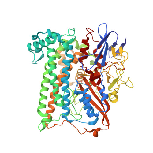
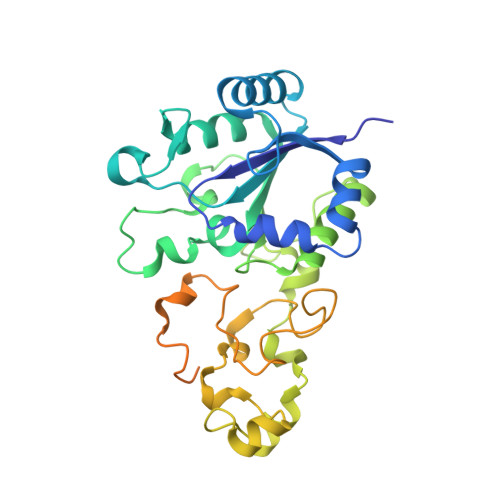
Mechanism of hydrogen activation by [NiFe] hydrogenases.

Evans, R.M.Brooke, E.J.Wehlin, S.A.Nomerotskaia, E.Sargent, F.Carr, S.B.Phillips, S.E.Armstrong, F.A.

(2016) Nat Chem Biol 12: 46-50

  • DOI: https://doi.org/10.1038/nchembio.1976
  • Primary Citation of Related Structures:  
    4UE3, 5A4F, 5A4I, 5A4M, 5ADU

  • PubMed Abstract: 

    The active site of [NiFe] hydrogenases contains a strictly conserved arginine that suspends a guanidine nitrogen atom <4.5 Å above the nickel and iron atoms. The guanidine headgroup interacts with the side chains of two conserved aspartic acid residues to complete an outer-shell canopy that has thus far proved intractable to investigation by site-directed mutagenesis. Using hydrogenase-1 from Escherichia coli, the strictly conserved residues R509 and D574 have been replaced by lysine (R509K) and asparagine (D574N) and the highly conserved D118 has been replaced by alanine (D118A) or asparagine (D118N/D574N). Each enzyme variant is stable, and their [(RS)2Niμ(SR)2Fe(CO)(CN)2] inner coordination shells are virtually unchanged. The R509K variant had >100-fold lower activity than native enzyme. Conversely, the variants D574N, D118A and D118N/D574N, in which the position of the guanidine headgroup is retained, showed 83%, 26% and 20% activity, respectively. The special kinetic requirement for R509 implicates the suspended guanidine group as the general base in H2 activation by [NiFe] hydrogenases.


  • Organizational Affiliation

    Department of Chemistry, University of Oxford, Oxford, UK.


Macromolecules
Find similar proteins by:  (by identity cutoff)  |  3D Structure
Entity ID: 1
MoleculeChains Sequence LengthOrganismDetailsImage
Hydrogenase-1 large chainA [auth L],
B [auth M]
582Escherichia coli str. K-12 substr. MC4100Mutation(s): 1 
Gene Names: hyaBb0973JW0955
EC: 1.12.99.6
UniProt
Find proteins for P0ACD8 (Escherichia coli (strain K12))
Explore P0ACD8 
Go to UniProtKB:  P0ACD8
Entity Groups  
Sequence Clusters30% Identity50% Identity70% Identity90% Identity95% Identity100% Identity
UniProt GroupP0ACD8
Sequence Annotations
Expand
  • Reference Sequence
Find similar proteins by:  (by identity cutoff)  |  3D Structure
Entity ID: 2
MoleculeChains Sequence LengthOrganismDetailsImage
Hydrogenase-1 small chainC [auth S],
D [auth T]
335Escherichia coli str. K-12 substr. MC4100Mutation(s): 0 
Gene Names: hyaAb0972JW0954
EC: 1.12.99.6
UniProt
Find proteins for P69739 (Escherichia coli (strain K12))
Explore P69739 
Go to UniProtKB:  P69739
Entity Groups  
Sequence Clusters30% Identity50% Identity70% Identity90% Identity95% Identity100% Identity
UniProt GroupP69739
Sequence Annotations
Expand
  • Reference Sequence
Small Molecules
Ligands 8 Unique
IDChains Name / Formula / InChI Key2D Diagram3D Interactions
SF4
Query on SF4

Download Ideal Coordinates CCD File 
L [auth S],
R [auth T]
IRON/SULFUR CLUSTER
Fe4 S4
LJBDFODJNLIPKO-UHFFFAOYSA-N
SF3
Query on SF3

Download Ideal Coordinates CCD File 
N [auth S],
T
FE4-S3 CLUSTER
Fe4 S3
QQACTBFBZNWJMV-UHFFFAOYSA-N
F3S
Query on F3S

Download Ideal Coordinates CCD File 
M [auth S],
S [auth T]
FE3-S4 CLUSTER
Fe3 S4
FCXHZBQOKRZXKS-UHFFFAOYSA-N
FCO
Query on FCO

Download Ideal Coordinates CCD File 
E [auth L],
H [auth M]
CARBONMONOXIDE-(DICYANO) IRON
C3 Fe N2 O
VBQUCMTXYFMTTE-UHFFFAOYSA-N
SO4
Query on SO4

Download Ideal Coordinates CCD File 
K [auth M],
Q [auth S]
SULFATE ION
O4 S
QAOWNCQODCNURD-UHFFFAOYSA-L
NI
Query on NI

Download Ideal Coordinates CCD File 
F [auth L],
I [auth M]
NICKEL (II) ION
Ni
VEQPNABPJHWNSG-UHFFFAOYSA-N
CL
Query on CL

Download Ideal Coordinates CCD File 
O [auth S],
P [auth S],
U [auth T],
V [auth T]
CHLORIDE ION
Cl
VEXZGXHMUGYJMC-UHFFFAOYSA-M
MG
Query on MG

Download Ideal Coordinates CCD File 
G [auth L],
J [auth M]
MAGNESIUM ION
Mg
JLVVSXFLKOJNIY-UHFFFAOYSA-N
Modified Residues  1 Unique
IDChains TypeFormula2D DiagramParent
CSO
Query on CSO
A [auth L],
B [auth M]
L-PEPTIDE LINKINGC3 H7 N O3 SCYS
Experimental Data & Validation

Experimental Data

  • Method: X-RAY DIFFRACTION
  • Resolution: 1.25 Å
  • R-Value Free: 0.157 
  • R-Value Work: 0.128 
  • R-Value Observed: 0.129 
  • Space Group: P 21 21 21
Unit Cell:
Length ( Å )Angle ( ˚ )
a = 93.757α = 90
b = 97.77β = 90
c = 183.288γ = 90
Software Package:
Software NamePurpose
REFMACrefinement
XDSdata reduction
XSCALEdata scaling
PHASERphasing

Structure Validation

View Full Validation Report


Entry History 

Deposition Data

Revision History  (Full details and data files)

  • Version 1.0: 2015-11-25
    Type: Initial release
  • Version 1.1: 2015-12-09
    Changes: Database references
  • Version 1.2: 2016-01-13
    Changes: Database references
  • Version 1.3: 2018-11-14
    Changes: Data collection, Database references, Derived calculations, Source and taxonomy, Structure summary
  • Version 1.4: 2024-01-10
    Changes: Data collection, Database references, Derived calculations, Other, Refinement description