5A1J

Periplasmic Binding Protein CeuE in complex with ferric 4-LICAM


Experimental Data Snapshot

  • Method: X-RAY DIFFRACTION
  • Resolution: 1.60 Å
  • R-Value Free: 0.312 
  • R-Value Work: 0.264 
  • R-Value Observed: 0.267 

wwPDB Validation   3D Report Full Report


Ligand Structure Quality Assessment 


This is version 1.1 of the entry. See complete history


Literature

Interactions of a Periplasmic Binding Protein with a Tetradentate Siderophore Mimic.

Raines, D.J.Moroz, O.V.Wilson, K.S.Duhme-Klair, A.

(2013) Angew Chem Int Ed Engl 52: 4595

  • DOI: https://doi.org/10.1002/anie.201300751
  • Primary Citation of Related Structures:  
    3ZKW, 5A1J

  • PubMed Abstract: 

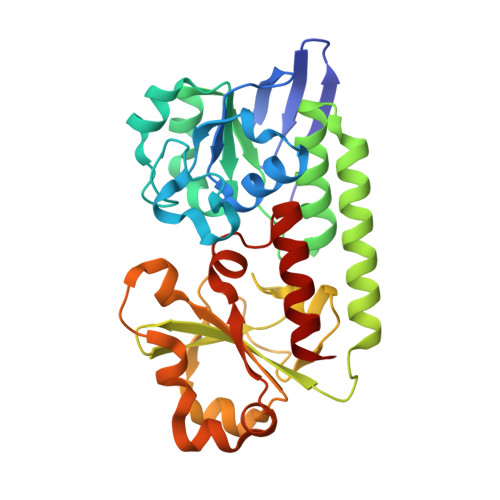
    Iron-bound structure: The ferric complex of a tetradentate siderophore mimic was synthesized and co-crystallized with the periplasmic binding protein CeuE of Campylobacter jejuni. In addition to electrostatic and hydrogen-bonding interactions between the binding pocket and the substrate, the structure showed direct coordination of two amino acid side chains to the Fe(III) center (orange, see figure).


  • Organizational Affiliation

    Department of Chemistry, University of York, Heslington, York, YO10 5DD, UK.


Macromolecules
Find similar proteins by:  (by identity cutoff)  |  3D Structure
Entity ID: 1
MoleculeChains Sequence LengthOrganismDetailsImage
ENTEROCHELIN UPTAKE PERIPLASMIC BINDING PROTEIN288Campylobacter jejuniMutation(s): 0 
UniProt
Find proteins for Q0P8Q4 (Campylobacter jejuni subsp. jejuni serotype O:2 (strain ATCC 700819 / NCTC 11168))
Explore Q0P8Q4 
Go to UniProtKB:  Q0P8Q4
Entity Groups  
Sequence Clusters30% Identity50% Identity70% Identity90% Identity95% Identity100% Identity
UniProt GroupQ0P8Q4
Sequence Annotations
Expand
  • Reference Sequence
Small Molecules
Ligands 2 Unique
IDChains Name / Formula / InChI Key2D Diagram3D Interactions
LCM
Query on LCM

Download Ideal Coordinates CCD File 
C [auth A]N,N'-butane-1,4-diylbis(2,3-dihydroxybenzamide)
C18 H20 N2 O6
FBRVRHJYPWFVCL-UHFFFAOYSA-N
FE
Query on FE

Download Ideal Coordinates CCD File 
B [auth A]FE (III) ION
Fe
VTLYFUHAOXGGBS-UHFFFAOYSA-N
Experimental Data & Validation

Experimental Data

  • Method: X-RAY DIFFRACTION
  • Resolution: 1.60 Å
  • R-Value Free: 0.312 
  • R-Value Work: 0.264 
  • R-Value Observed: 0.267 
  • Space Group: P 21 21 21
Unit Cell:
Length ( Å )Angle ( ˚ )
a = 60.42α = 90
b = 66.89β = 90
c = 67.3γ = 90
Software Package:
Software NamePurpose
REFMACrefinement
xia2data reduction
MOLREPphasing

Structure Validation

View Full Validation Report


Ligand Structure Quality Assessment 


Entry History 

Deposition Data

Revision History  (Full details and data files)

  • Version 1.0: 2015-05-13
    Type: Initial release
  • Version 1.1: 2024-01-10
    Changes: Data collection, Database references, Derived calculations, Other, Refinement description