4ZHX

Novel binding site for allosteric activation of AMPK


Experimental Data Snapshot

  • Method: X-RAY DIFFRACTION
  • Resolution: 2.99 Å
  • R-Value Free: 0.243 
  • R-Value Work: 0.225 
  • R-Value Observed: 0.226 

wwPDB Validation   3D Report Full Report


This is version 1.3 of the entry. See complete history


Literature

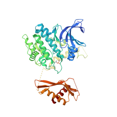
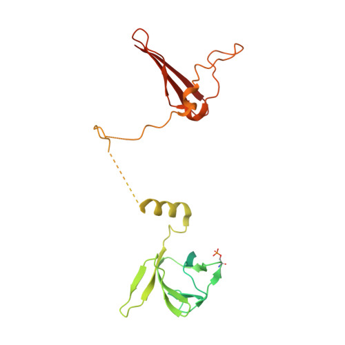
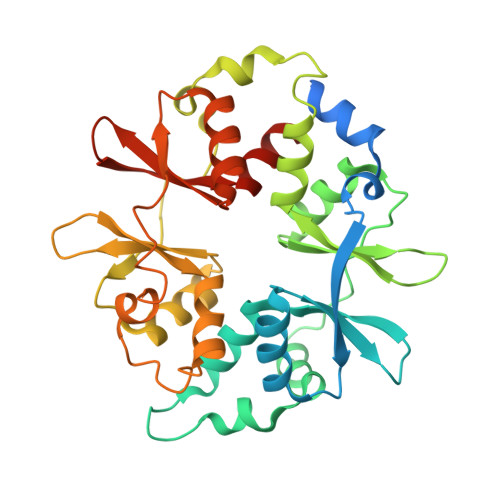
Structural basis of allosteric and synergistic activation of AMPK by furan-2-phosphonic derivative C2 binding.

Langendorf, C.G.Ngoei, K.R.Scott, J.W.Ling, N.X.Issa, S.M.Gorman, M.A.Parker, M.W.Sakamoto, K.Oakhill, J.S.Kemp, B.E.

(2016) Nat Commun 7: 10912-10912

  • DOI: https://doi.org/10.1038/ncomms10912
  • Primary Citation of Related Structures:  
    4ZHX, 5EZV

  • PubMed Abstract: 

    The metabolic stress-sensing enzyme AMP-activated protein kinase (AMPK) is responsible for regulating metabolism in response to energy supply and demand. Drugs that activate AMPK may be useful in the treatment of metabolic diseases including type 2 diabetes. We have determined the crystal structure of AMPK in complex with its activator 5-(5-hydroxyl-isoxazol-3-yl)-furan-2-phosphonic acid (C2), revealing two C2-binding sites in the γ-subunit distinct from nucleotide sites. C2 acts synergistically with the drug A769662 to activate AMPK α1-containing complexes independent of upstream kinases. Our results show that dual drug therapies could be effective AMPK-targeting strategies to treat metabolic diseases.


  • Organizational Affiliation

    Protein Chemistry &Metabolism, St Vincent's Institute of Medical Research, University of Melbourne, 41 Victoria Parade, Fitzroy, Victoria 3065, Australia.


Macromolecules
Find similar proteins by:  (by identity cutoff)  |  3D Structure
Entity ID: 1
MoleculeChains Sequence LengthOrganismDetailsImage
5'-AMP-activated protein kinase catalytic subunit alpha-2
A, C
565Homo sapiensMutation(s): 1 
Gene Names: PRKAA2AMPKAMPK2
EC: 2.7.11.1 (PDB Primary Data), 2.7.11.27 (PDB Primary Data), 2.7.11.31 (PDB Primary Data)
UniProt & NIH Common Fund Data Resources
Find proteins for P54646 (Homo sapiens)
Explore P54646 
Go to UniProtKB:  P54646
PHAROS:  P54646
GTEx:  ENSG00000162409 
Entity Groups  
Sequence Clusters30% Identity50% Identity70% Identity90% Identity95% Identity100% Identity
UniProt GroupP54646
Sequence Annotations
Expand
  • Reference Sequence
Find similar proteins by:  (by identity cutoff)  |  3D Structure
Entity ID: 2
MoleculeChains Sequence LengthOrganismDetailsImage
5'-AMP-activated protein kinase subunit beta-1
B, D
270Homo sapiensMutation(s): 0 
Gene Names: PRKAB1AMPK
UniProt & NIH Common Fund Data Resources
Find proteins for Q9Y478 (Homo sapiens)
Explore Q9Y478 
Go to UniProtKB:  Q9Y478
PHAROS:  Q9Y478
GTEx:  ENSG00000111725 
Entity Groups  
Sequence Clusters30% Identity50% Identity70% Identity90% Identity95% Identity100% Identity
UniProt GroupQ9Y478
Sequence Annotations
Expand
  • Reference Sequence
Find similar proteins by:  (by identity cutoff)  |  3D Structure
Entity ID: 3
MoleculeChains Sequence LengthOrganismDetailsImage
5'-AMP-activated protein kinase subunit gamma-1
E, F
336Homo sapiensMutation(s): 0 
Gene Names: PRKAG1
UniProt & NIH Common Fund Data Resources
Find proteins for P54619 (Homo sapiens)
Explore P54619 
Go to UniProtKB:  P54619
PHAROS:  P54619
GTEx:  ENSG00000181929 
Entity Groups  
Sequence Clusters30% Identity50% Identity70% Identity90% Identity95% Identity100% Identity
UniProt GroupP54619
Sequence Annotations
Expand
  • Reference Sequence
Small Molecules
Ligands 4 Unique
IDChains Name / Formula / InChI Key2D Diagram3D Interactions
4O7
Query on 4O7

Download Ideal Coordinates CCD File 
G [auth A],
I [auth C]
(5S,6R,7R,9R,13cR,14R,16aS)-6-methoxy-5-methyl-7-(methylamino)-6,7,8,9,14,15,16,16a-octahydro-5H,13cH-5,9-epoxy-4b,9a,1 5-triazadibenzo[b,h]cyclonona[1,2,3,4-jkl]cyclopenta[e]-as-indacen-14-ol
C28 H30 N4 O3
FJJCTPAMIAOFJX-VWKRKTLRSA-N
C1V
Query on C1V

Download Ideal Coordinates CCD File 
H [auth B],
J [auth C]
3-[4-(2-hydroxyphenyl)phenyl]-4-oxidanyl-6-oxidanylidene-7H-thieno[2,3-b]pyridine-5-carbonitrile
C20 H12 N2 O3 S
CTESJDQKVOEUOY-UHFFFAOYSA-N
AMP
Query on AMP

Download Ideal Coordinates CCD File 
M [auth F],
N [auth F]
ADENOSINE MONOPHOSPHATE
C10 H14 N5 O7 P
UDMBCSSLTHHNCD-KQYNXXCUSA-N
C2Z
Query on C2Z

Download Ideal Coordinates CCD File 
K [auth E],
L [auth E]
5-(5-hydroxyl-isoxazol-3-yl)-furan-2-phosphonic acid
C7 H6 N O6 P
BRLJHMCTDKAUAY-UHFFFAOYSA-N
Modified Residues  2 Unique
IDChains TypeFormula2D DiagramParent
TPO
Query on TPO
A, C
L-PEPTIDE LINKINGC4 H10 N O6 PTHR
SEP
Query on SEP
B, D
L-PEPTIDE LINKINGC3 H8 N O6 PSER
Binding Affinity Annotations 
IDSourceBinding Affinity
C1V BindingDB:  4ZHX Kd: min: 487, max: 1.45e+4 (nM) from 3 assay(s)
EC50: 800 (nM) from 1 assay(s)
Experimental Data & Validation

Experimental Data

  • Method: X-RAY DIFFRACTION
  • Resolution: 2.99 Å
  • R-Value Free: 0.243 
  • R-Value Work: 0.225 
  • R-Value Observed: 0.226 
  • Space Group: P 1 21 1
Unit Cell:
Length ( Å )Angle ( ˚ )
a = 75.95α = 90
b = 134.24β = 93.02
c = 141.48γ = 90
Software Package:
Software NamePurpose
BUSTER-TNTrefinement
Aimlessdata scaling
PDB_EXTRACTdata extraction
XDSdata reduction
PHASERphasing

Structure Validation

View Full Validation Report


Entry History 

Deposition Data

Revision History  (Full details and data files)

  • Version 1.0: 2016-03-09
    Type: Initial release
  • Version 1.1: 2016-03-23
    Changes: Database references
  • Version 1.2: 2017-09-27
    Changes: Author supporting evidence, Data collection, Derived calculations
  • Version 1.3: 2023-09-27
    Changes: Data collection, Database references, Derived calculations, Refinement description, Structure summary