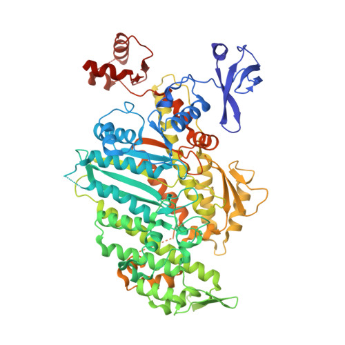
4ZG4

Myosin Vc Pre-powerstroke


Experimental Data Snapshot

  • Method: X-RAY DIFFRACTION
  • Resolution: 2.36 Å
  • R-Value Free: 0.213 
  • R-Value Work: 0.173 
  • R-Value Observed: 0.175 

wwPDB Validation   3D Report Full Report


Ligand Structure Quality Assessment 


This is version 1.4 of the entry. See complete history


Literature

Force-producing ADP state of myosin bound to actin.

Wulf, S.F.Ropars, V.Fujita-Becker, S.Oster, M.Hofhaus, G.Trabuco, L.G.Pylypenko, O.Sweeney, H.L.Houdusse, A.M.Schroder, R.R.

(2016) Proc Natl Acad Sci U S A 113: E1844-E1852

  • DOI: https://doi.org/10.1073/pnas.1516598113
  • Primary Citation of Related Structures:  
    4ZG4

  • PubMed Abstract: 

    Molecular motors produce force when they interact with their cellular tracks. For myosin motors, the primary force-generating state has MgADP tightly bound, whereas myosin is strongly bound to actin. We have generated an 8-Å cryoEM reconstruction of this state for myosin V and used molecular dynamics flexed fitting for model building. We compare this state to the subsequent state on actin (Rigor). The ADP-bound structure reveals that the actin-binding cleft is closed, even though MgADP is tightly bound. This state is accomplished by a previously unseen conformation of the β-sheet underlying the nucleotide pocket. The transition from the force-generating ADP state to Rigor requires a 9.5° rotation of the myosin lever arm, coupled to a β-sheet rearrangement. Thus, the structure reveals the detailed rearrangements underlying myosin force generation as well as the basis of strain-dependent ADP release that is essential for processive myosins, such as myosin V.


  • Organizational Affiliation

    Cryo Electron Microscopy, CellNetworks, BioQuant, Universitätsklinikum Heidelberg, 69120 Heidelberg, Germany;


Macromolecules
Find similar proteins by:  (by identity cutoff)  |  3D Structure
Entity ID: 1
MoleculeChains Sequence LengthOrganismDetailsImage
myosin-VcA [auth B],
B [auth E]
764Homo sapiensMutation(s): 0 
Gene Names: MYO5C
UniProt & NIH Common Fund Data Resources
Find proteins for Q9NQX4 (Homo sapiens)
Explore Q9NQX4 
Go to UniProtKB:  Q9NQX4
PHAROS:  Q9NQX4
GTEx:  ENSG00000128833 
Entity Groups  
Sequence Clusters30% Identity50% Identity70% Identity90% Identity95% Identity100% Identity
UniProt GroupQ9NQX4
Sequence Annotations
Expand
  • Reference Sequence
Small Molecules
Ligands 4 Unique
IDChains Name / Formula / InChI Key2D Diagram3D Interactions
ADP
Query on ADP

Download Ideal Coordinates CCD File 
E [auth B],
I [auth E]
ADENOSINE-5'-DIPHOSPHATE
C10 H15 N5 O10 P2
XTWYTFMLZFPYCI-KQYNXXCUSA-N
VO4
Query on VO4

Download Ideal Coordinates CCD File 
D [auth B],
H [auth E]
VANADATE ION
O4 V
LSGOVYNHVSXFFJ-UHFFFAOYSA-N
EDO
Query on EDO

Download Ideal Coordinates CCD File 
F [auth B]1,2-ETHANEDIOL
C2 H6 O2
LYCAIKOWRPUZTN-UHFFFAOYSA-N
MG
Query on MG

Download Ideal Coordinates CCD File 
C [auth B],
G [auth E]
MAGNESIUM ION
Mg
JLVVSXFLKOJNIY-UHFFFAOYSA-N
Experimental Data & Validation

Experimental Data

  • Method: X-RAY DIFFRACTION
  • Resolution: 2.36 Å
  • R-Value Free: 0.213 
  • R-Value Work: 0.173 
  • R-Value Observed: 0.175 
  • Space Group: P 1 21 1
Unit Cell:
Length ( Å )Angle ( ˚ )
a = 118.28α = 90
b = 67.32β = 108.48
c = 133.18γ = 90
Software Package:
Software NamePurpose
PHENIXrefinement
XDSdata reduction
XSCALEdata scaling
MOLREPphasing

Structure Validation

View Full Validation Report


Ligand Structure Quality Assessment 


Entry History & Funding Information

Deposition Data


Funding OrganizationLocationGrant Number
French National Research AgencyFranceANR-13-BSV8-0019-01

Revision History  (Full details and data files)

  • Version 1.0: 2016-03-23
    Type: Initial release
  • Version 1.1: 2016-03-30
    Changes: Database references
  • Version 1.2: 2016-04-13
    Changes: Database references
  • Version 1.3: 2017-09-06
    Changes: Author supporting evidence
  • Version 1.4: 2024-01-10
    Changes: Data collection, Database references, Derived calculations, Refinement description