4ZB0

A dehydrated form of glucose isomerase collected at room temperature.


Experimental Data Snapshot

  • Method: X-RAY DIFFRACTION
  • Resolution: 2.00 Å
  • R-Value Free: 0.180 
  • R-Value Work: 0.137 
  • R-Value Observed: 0.139 

wwPDB Validation   3D Report Full Report


Ligand Structure Quality Assessment 


This is version 1.3 of the entry. See complete history


Literature

A generic protocol for protein crystal dehydration using the HC1b humidity controller.

Lobley, C.M.Sandy, J.Sanchez-Weatherby, J.Mazzorana, M.Krojer, T.Nowak, R.P.Sorensen, T.L.

(2016) Acta Crystallogr D Struct Biol 72: 629-640

  • DOI: https://doi.org/10.1107/S2059798316003065
  • Primary Citation of Related Structures:  
    4ZB0, 4ZB2, 4ZB5, 4ZBC

  • PubMed Abstract: 

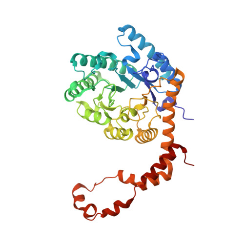
    Dehydration may change the crystal lattice and affect the mosaicity, resolution and quality of X-ray diffraction data. A dehydrating environment can be generated around a crystal in several ways with various degrees of precision and complexity. This study uses a high-precision crystal humidifier/dehumidifier to provide an airstream of known relative humidity in which the crystals are mounted: a precise yet hassle-free approach to altering crystal hydration. A protocol is introduced to assess the impact of crystal dehydration systematically applied to nine experimental crystal systems. In one case, that of glucose isomerase, dehydration triggering a change of space group from I222 to P21212 was observed. This observation is supported by an extended study of the behaviour of the glucose isomerase crystal structure during crystal dehydration.


  • Organizational Affiliation

    Diamond Light Source, Harwell Science and Innovation Campus, Didcot OX11 0DE, England.


Macromolecules
Find similar proteins by:  (by identity cutoff)  |  3D Structure
Entity ID: 1
MoleculeChains Sequence LengthOrganismDetailsImage
Xylose isomerase
A, B
387Streptomyces rubiginosusMutation(s): 0 
Gene Names: xylA
EC: 5.3.1.5
UniProt
Find proteins for P24300 (Streptomyces rubiginosus)
Explore P24300 
Go to UniProtKB:  P24300
Entity Groups  
Sequence Clusters30% Identity50% Identity70% Identity90% Identity95% Identity100% Identity
UniProt GroupP24300
Sequence Annotations
Expand
  • Reference Sequence
Small Molecules
Ligands 3 Unique
IDChains Name / Formula / InChI Key2D Diagram3D Interactions
FRU
Query on FRU

Download Ideal Coordinates CCD File 
I [auth A],
P [auth B]
beta-D-fructofuranose
C6 H12 O6
RFSUNEUAIZKAJO-ARQDHWQXSA-N
GLC
Query on GLC

Download Ideal Coordinates CCD File 
H [auth A],
O [auth B]
alpha-D-glucopyranose
C6 H12 O6
WQZGKKKJIJFFOK-DVKNGEFBSA-N
MN
Query on MN

Download Ideal Coordinates CCD File 
C [auth A]
D [auth A]
E [auth A]
F [auth A]
G [auth A]
C [auth A],
D [auth A],
E [auth A],
F [auth A],
G [auth A],
J [auth B],
K [auth B],
L [auth B],
M [auth B],
N [auth B]
MANGANESE (II) ION
Mn
WAEMQWOKJMHJLA-UHFFFAOYSA-N
Experimental Data & Validation

Experimental Data

  • Method: X-RAY DIFFRACTION
  • Resolution: 2.00 Å
  • R-Value Free: 0.180 
  • R-Value Work: 0.137 
  • R-Value Observed: 0.139 
  • Space Group: P 21 21 2
Unit Cell:
Length ( Å )Angle ( ˚ )
a = 83.485α = 90
b = 95.02β = 90
c = 98.484γ = 90
Software Package:
Software NamePurpose
REFMACrefinement
xia2data reduction
xia2data scaling
MOLREPphasing
DIALSdata reduction
DIALSdata scaling

Structure Validation

View Full Validation Report


Ligand Structure Quality Assessment 


Entry History 

Deposition Data

  • Released Date: 2016-03-23 
  • Deposition Author(s): Sandy, J.

Revision History  (Full details and data files)

  • Version 1.0: 2016-03-23
    Type: Initial release
  • Version 1.1: 2016-05-11
    Changes: Database references
  • Version 1.2: 2020-07-29
    Type: Remediation
    Reason: Carbohydrate remediation
    Changes: Data collection, Derived calculations, Structure summary
  • Version 1.3: 2024-01-10
    Changes: Data collection, Database references, Refinement description, Structure summary