4XGY

GFP based antibody (fluorobody)


Experimental Data Snapshot

  • Method: X-RAY DIFFRACTION
  • Resolution: 1.49 Å
  • R-Value Free: 0.167 
  • R-Value Work: 0.145 
  • R-Value Observed: 0.146 

wwPDB Validation   3D Report Full Report


This is version 2.0 of the entry. See complete history


Literature

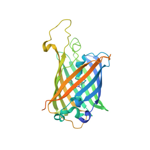
The structure of a GFP-based antibody (fluorobody) to TLH, a toxin from Vibrio parahaemolyticus.

Chen, Y.Huang, X.Wang, R.Wang, S.Shi, N.

(2015) Acta Crystallogr F Struct Biol Commun 71: 913-918

  • DOI: https://doi.org/10.1107/S2053230X15008845
  • Primary Citation of Related Structures:  
    4XGY

  • PubMed Abstract: 

    A fluorobody is a manmade hybrid molecule that is composed of green fluorescent protein (GFP) and a fragment of antibody, which combines the affinity and specificity of an antibody with the visibility of a GFP. It is able to provide a real-time indication of binding while avoiding the use of tags and secondary binding reagents. Here, the expression, purification and crystal structure of a recombinant fluorobody for TLH (thermolabile haemolysin), a toxin from the lethal food-borne disease bacterium Vibrio parahaemolyticus, are presented. This is the first structure of a fluorobody to be reported. Crystals belonging to space group P4(3)2(1)2, with unit-cell parameters a = b = 63.35, c = 125.90 Å, were obtained by vapour diffusion in hanging drops and the structure was refined to an Rfree of 16.7% at 1.5 Å resolution. The structure shows a CDR loop of the antibody on the GFP scaffold.


  • Organizational Affiliation

    State Key Laboratory of Structural Chemistry, Fujian Institute of Research on the Structure of Matter, Chinese Academy of Sciences, 155 Yangqiao Road West, Fuzhou 350002, People's Republic of China.


Macromolecules
Find similar proteins by:  (by identity cutoff)  |  3D Structure
Entity ID: 1
MoleculeChains Sequence LengthOrganismDetailsImage
Green fluorescent protein, mAb LCDR3, Green fluorescent protein303Aequorea victoriaVibrio parahaemolyticussynthetic constructMutation(s): 11 
Gene Names: GFP
UniProt
Find proteins for A0A059PIQ0 (Aequorea victoria)
Explore A0A059PIQ0 
Go to UniProtKB:  A0A059PIQ0
Entity Groups  
Sequence Clusters30% Identity50% Identity70% Identity90% Identity95% Identity100% Identity
UniProt GroupA0A059PIQ0
Sequence Annotations
Expand
  • Reference Sequence
Small Molecules
Ligands 1 Unique
IDChains Name / Formula / InChI Key2D Diagram3D Interactions
PE5
Query on PE5

Download Ideal Coordinates CCD File 
B [auth A],
C [auth A]
3,6,9,12,15,18,21,24-OCTAOXAHEXACOSAN-1-OL
C18 H38 O9
CUDPPTPIUWYGFI-UHFFFAOYSA-N
Modified Residues  1 Unique
IDChains TypeFormula2D DiagramParent
CRO
Query on CRO
A
L-PEPTIDE LINKINGC15 H17 N3 O5THR, TYR, GLY
Experimental Data & Validation

Experimental Data

  • Method: X-RAY DIFFRACTION
  • Resolution: 1.49 Å
  • R-Value Free: 0.167 
  • R-Value Work: 0.145 
  • R-Value Observed: 0.146 
  • Space Group: P 43 21 2
Unit Cell:
Length ( Å )Angle ( ˚ )
a = 63.35α = 90
b = 63.35β = 90
c = 125.9γ = 90
Software Package:
Software NamePurpose
PHENIXrefinement
iMOSFLMdata reduction
iMOSFLMdata scaling
PHASERphasing

Structure Validation

View Full Validation Report


Entry History 

Deposition Data

Revision History  (Full details and data files)

  • Version 1.0: 2015-07-01
    Type: Initial release
  • Version 1.1: 2015-07-22
    Changes: Database references
  • Version 1.2: 2023-11-08
    Changes: Advisory, Data collection, Database references, Derived calculations, Refinement description, Source and taxonomy, Structure summary
  • Version 2.0: 2023-11-15
    Changes: Atomic model, Data collection