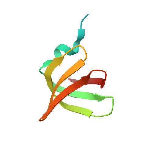
4X9D

High-resolution structure of Hfq from Methanococcus jannaschii in complex with UMP


Experimental Data Snapshot

  • Method: X-RAY DIFFRACTION
  • Resolution: 1.50 Å
  • R-Value Free: 0.203 
  • R-Value Work: 0.174 
  • R-Value Observed: 0.175 

wwPDB Validation   3D Report Full Report


Ligand Structure Quality Assessment 


This is version 1.3 of the entry. See complete history


Literature

Characterization of RNA-binding properties of the archaeal Hfq-like protein from Methanococcus jannaschii.

Nikulin, A.Mikhailina, A.Lekontseva, N.Balobanov, V.Nikonova, E.Tishchenko, S.

(2017) J Biomol Struct Dyn 35: 1615-1628

  • DOI: https://doi.org/10.1080/07391102.2016.1189849
  • Primary Citation of Related Structures:  
    4X9C, 4X9D, 5DY9

  • PubMed Abstract: 

    The Sm and Sm-like proteins are widely distributed among bacteria, archaea and eukarya. They participate in many processes related to RNA-processing and regulation of gene expression. While the function of the bacterial Lsm protein Hfq and eukaryotic Sm/Lsm proteins is rather well studied, the role of Lsm proteins in Archaea is investigated poorly. In this work, the RNA-binding ability of an archaeal Hfq-like protein from Methanococcus jannaschii has been studied by X-ray crystallography, anisotropy fluorescence and surface plasmon resonance. It has been found that MjaHfq preserves the proximal RNA-binding site that usually recognizes uridine-rich sequences. Distal adenine-binding and lateral RNA-binding sites show considerable structural changes as compared to bacterial Hfq. MjaHfq did not bind mononucleotides at these sites and would not recognize single-stranded RNA as its bacterial homologues. Nevertheless, MjaHfq possesses affinity to poly(A) RNA that seems to bind at the unstructured positive-charged N-terminal tail of the protein.


  • Organizational Affiliation

    a Institute of Protein Research , Russian Academy of Sciences , Pushchino , Moscow region , 142290 , Russia.


Macromolecules
Find similar proteins by:  (by identity cutoff)  |  3D Structure
Entity ID: 1
MoleculeChains Sequence LengthOrganismDetailsImage
Uncharacterized protein MJ1435
A, B, C, D, E
A, B, C, D, E, F
71Methanocaldococcus jannaschiiMutation(s): 0 
Gene Names: MJ1435
UniProt
Find proteins for Q58830 (Methanocaldococcus jannaschii (strain ATCC 43067 / DSM 2661 / JAL-1 / JCM 10045 / NBRC 100440))
Explore Q58830 
Go to UniProtKB:  Q58830
Entity Groups  
Sequence Clusters30% Identity50% Identity70% Identity90% Identity95% Identity100% Identity
UniProt GroupQ58830
Sequence Annotations
Expand
  • Reference Sequence
Small Molecules
Ligands 8 Unique
IDChains Name / Formula / InChI Key2D Diagram3D Interactions
U5P
Query on U5P

Download Ideal Coordinates CCD File 
AA [auth F]
J [auth A]
N [auth B]
R [auth C]
S [auth D]
AA [auth F],
J [auth A],
N [auth B],
R [auth C],
S [auth D],
W [auth E]
URIDINE-5'-MONOPHOSPHATE
C9 H13 N2 O9 P
DJJCXFVJDGTHFX-XVFCMESISA-N
PG4
Query on PG4

Download Ideal Coordinates CCD File 
O [auth C],
T [auth E]
TETRAETHYLENE GLYCOL
C8 H18 O5
UWHCKJMYHZGTIT-UHFFFAOYSA-N
PGE
Query on PGE

Download Ideal Coordinates CCD File 
G [auth A],
U [auth E]
TRIETHYLENE GLYCOL
C6 H14 O4
ZIBGPFATKBEMQZ-UHFFFAOYSA-N
PEG
Query on PEG

Download Ideal Coordinates CCD File 
X [auth F]DI(HYDROXYETHYL)ETHER
C4 H10 O3
MTHSVFCYNBDYFN-UHFFFAOYSA-N
SO4
Query on SO4

Download Ideal Coordinates CCD File 
H [auth A],
Y [auth F]
SULFATE ION
O4 S
QAOWNCQODCNURD-UHFFFAOYSA-L
EDO
Query on EDO

Download Ideal Coordinates CCD File 
K [auth B]1,2-ETHANEDIOL
C2 H6 O2
LYCAIKOWRPUZTN-UHFFFAOYSA-N
CL
Query on CL

Download Ideal Coordinates CCD File 
Q [auth C],
V [auth E]
CHLORIDE ION
Cl
VEXZGXHMUGYJMC-UHFFFAOYSA-M
NA
Query on NA

Download Ideal Coordinates CCD File 
I [auth A],
L [auth B],
M [auth B],
P [auth C],
Z [auth F]
SODIUM ION
Na
FKNQFGJONOIPTF-UHFFFAOYSA-N
Experimental Data & Validation

Experimental Data

  • Method: X-RAY DIFFRACTION
  • Resolution: 1.50 Å
  • R-Value Free: 0.203 
  • R-Value Work: 0.174 
  • R-Value Observed: 0.175 
  • Space Group: P 21 21 21
Unit Cell:
Length ( Å )Angle ( ˚ )
a = 57.741α = 90
b = 66.874β = 90
c = 109.31γ = 90
Software Package:
Software NamePurpose
PHENIXrefinement
XDSdata reduction
SCALAdata scaling
PHASERphasing

Structure Validation

View Full Validation Report


Ligand Structure Quality Assessment 


Entry History & Funding Information

Deposition Data


Funding OrganizationLocationGrant Number
Russian Scientific FoundationRussian Federation14-14-00496

Revision History  (Full details and data files)

  • Version 1.0: 2015-12-23
    Type: Initial release
  • Version 1.1: 2017-02-22
    Changes: Database references
  • Version 1.2: 2017-05-24
    Changes: Database references
  • Version 1.3: 2024-01-10
    Changes: Data collection, Database references, Derived calculations, Refinement description