4WZA

Asymmetric Nucleotide Binding in the Nitrogenase Complex


Experimental Data Snapshot

  • Method: X-RAY DIFFRACTION
  • Resolution: 1.90 Å
  • R-Value Free: 0.187 
  • R-Value Work: 0.145 
  • R-Value Observed: 0.147 

wwPDB Validation   3D Report Full Report


Ligand Structure Quality Assessment 


This is version 1.5 of the entry. See complete history


Literature

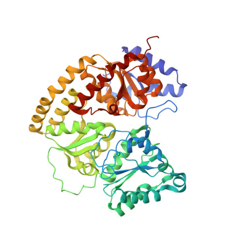
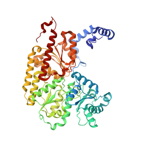
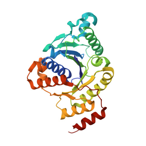
Structural evidence for asymmetrical nucleotide interactions in nitrogenase.

Tezcan, F.A.Kaiser, J.T.Howard, J.B.Rees, D.C.

(2015) J Am Chem Soc 137: 146-149

  • DOI: https://doi.org/10.1021/ja511945e
  • Primary Citation of Related Structures:  
    4WZA

  • PubMed Abstract: 

    The roles of ATP hydrolysis in electron-transfer (ET) reactions of the nitrogenase catalytic cycle remain obscure. Here, we present a new structure of a nitrogenase complex crystallized with MgADP and MgAMPPCP, an ATP analogue. In this structure the two nucleotides are bound asymmetrically by the Fe-protein subunits connected to the two different MoFe-protein subunits. This binding mode suggests that ATP hydrolysis and phosphate release may proceed by a stepwise mechanism. Through the associated Fe-protein conformational changes, a stepwise mechanism is anticipated to prolong the lifetime of the Fe-protein-MoFe-protein complex and, in turn, could orchestrate the sequence of intracomplex ET required for substrate reduction.


  • Organizational Affiliation

    Department of Chemistry and Biochemistry, University of California, San Diego , La Jolla, California 92093-0356, United States.


Macromolecules
Find similar proteins by:  (by identity cutoff)  |  3D Structure
Entity ID: 1
MoleculeChains Sequence LengthOrganismDetailsImage
Nitrogenase molybdenum-iron protein alpha chain
A, C
477Azotobacter vinelandiiMutation(s): 1 
EC: 1.18.6.1
UniProt
Find proteins for P07328 (Azotobacter vinelandii)
Explore P07328 
Go to UniProtKB:  P07328
Entity Groups  
Sequence Clusters30% Identity50% Identity70% Identity90% Identity95% Identity100% Identity
UniProt GroupP07328
Sequence Annotations
Expand
  • Reference Sequence
Find similar proteins by:  (by identity cutoff)  |  3D Structure
Entity ID: 2
MoleculeChains Sequence LengthOrganismDetailsImage
Nitrogenase molybdenum-iron protein beta chain
B, D
522Azotobacter vinelandiiMutation(s): 0 
EC: 1.18.6.1
UniProt
Find proteins for P07329 (Azotobacter vinelandii)
Explore P07329 
Go to UniProtKB:  P07329
Entity Groups  
Sequence Clusters30% Identity50% Identity70% Identity90% Identity95% Identity100% Identity
UniProt GroupP07329
Sequence Annotations
Expand
  • Reference Sequence
Find similar proteins by:  (by identity cutoff)  |  3D Structure
Entity ID: 3
MoleculeChains Sequence LengthOrganismDetailsImage
Nitrogenase iron protein 1
E, F, G, H
276Azotobacter vinelandiiMutation(s): 0 
EC: 1.18.6.1
UniProt
Find proteins for P00459 (Azotobacter vinelandii)
Explore P00459 
Go to UniProtKB:  P00459
Entity Groups  
Sequence Clusters30% Identity50% Identity70% Identity90% Identity95% Identity100% Identity
UniProt GroupP00459
Sequence Annotations
Expand
  • Reference Sequence
Small Molecules
Ligands 8 Unique
IDChains Name / Formula / InChI Key2D Diagram3D Interactions
ICS
Query on ICS

Download Ideal Coordinates CCD File 
J [auth A],
N [auth C]
iron-sulfur-molybdenum cluster with interstitial carbon
C Fe7 Mo S9
DDQFAOMIVKLFON-UHFFFAOYSA-N
CLF
Query on CLF

Download Ideal Coordinates CCD File 
K [auth A],
O [auth C]
FE(8)-S(7) CLUSTER
Fe8 S7
JKVMXLBGZBULKV-UHFFFAOYSA-N
ACP
Query on ACP

Download Ideal Coordinates CCD File 
U [auth F],
Z [auth H]
PHOSPHOMETHYLPHOSPHONIC ACID ADENYLATE ESTER
C11 H18 N5 O12 P3
UFZTZBNSLXELAL-IOSLPCCCSA-N
ADP
Query on ADP

Download Ideal Coordinates CCD File 
S [auth E],
X [auth G]
ADENOSINE-5'-DIPHOSPHATE
C10 H15 N5 O10 P2
XTWYTFMLZFPYCI-KQYNXXCUSA-N
SF4
Query on SF4

Download Ideal Coordinates CCD File 
Q [auth E],
V [auth G]
IRON/SULFUR CLUSTER
Fe4 S4
LJBDFODJNLIPKO-UHFFFAOYSA-N
HCA
Query on HCA

Download Ideal Coordinates CCD File 
I [auth A],
M [auth C]
3-HYDROXY-3-CARBOXY-ADIPIC ACID
C7 H10 O7
XKJVEVRQMLKSMO-SSDOTTSWSA-N
FE
Query on FE

Download Ideal Coordinates CCD File 
L [auth B],
P [auth D]
FE (III) ION
Fe
VTLYFUHAOXGGBS-UHFFFAOYSA-N
MG
Query on MG

Download Ideal Coordinates CCD File 
R [auth E],
T [auth F],
W [auth G],
Y [auth H]
MAGNESIUM ION
Mg
JLVVSXFLKOJNIY-UHFFFAOYSA-N
Experimental Data & Validation

Experimental Data

  • Method: X-RAY DIFFRACTION
  • Resolution: 1.90 Å
  • R-Value Free: 0.187 
  • R-Value Work: 0.145 
  • R-Value Observed: 0.147 
  • Space Group: P 21 21 21
Unit Cell:
Length ( Å )Angle ( ˚ )
a = 110.201α = 90
b = 120.412β = 90
c = 264.318γ = 90
Software Package:
Software NamePurpose
PHENIXrefinement
Aimlessdata scaling
PDB_EXTRACTdata extraction
MOLREPphasing

Structure Validation

View Full Validation Report


Ligand Structure Quality Assessment 


Entry History & Funding Information

Deposition Data


Funding OrganizationLocationGrant Number
National Institutes of Health/National Institute of General Medical Sciences (NIH/NIGMS)United StatesGM099813
National Institutes of Health/National Institute of General Medical Sciences (NIH/NIGMS)United StatesGM045162

Revision History  (Full details and data files)

  • Version 1.0: 2014-12-31
    Type: Initial release
  • Version 1.1: 2015-01-28
    Changes: Database references
  • Version 1.2: 2017-09-06
    Changes: Advisory, Author supporting evidence, Derived calculations, Refinement description, Source and taxonomy
  • Version 1.3: 2017-11-22
    Changes: Refinement description
  • Version 1.4: 2019-12-25
    Changes: Author supporting evidence
  • Version 1.5: 2023-09-27
    Changes: Advisory, Data collection, Database references, Derived calculations, Refinement description