4WKU

n-Alkylboronic Acid Inhibitors Reveal Determinants of Ligand Specificity in the Quorum-Quenching and Siderophore Biosynthetic Enzyme PvdQ


Experimental Data Snapshot

  • Method: X-RAY DIFFRACTION
  • Resolution: 2.00 Å
  • R-Value Free: 0.215 
  • R-Value Work: 0.180 
  • R-Value Observed: 0.182 

wwPDB Validation   3D Report Full Report


This is version 1.1 of the entry. See complete history


Literature

n-Alkylboronic Acid Inhibitors Reveal Determinants of Ligand Specificity in the Quorum-Quenching and Siderophore Biosynthetic Enzyme PvdQ.

Clevenger, K.D.Wu, R.Liu, D.Fast, W.

(2014) Biochemistry 53: 6679-6686

  • DOI: https://doi.org/10.1021/bi501086s
  • Primary Citation of Related Structures:  
    4WKS, 4WKT, 4WKU, 4WKV

  • PubMed Abstract: 

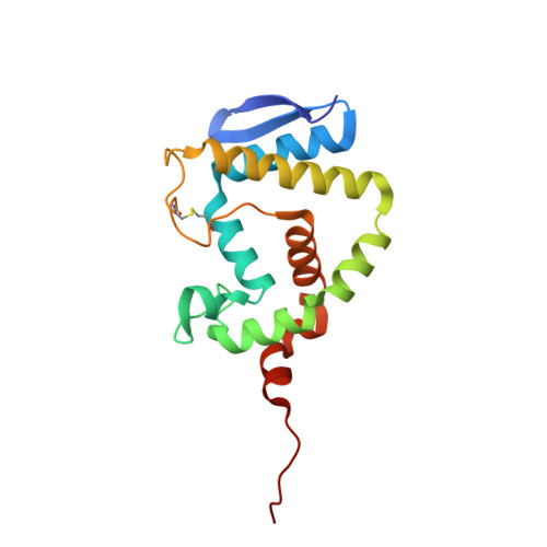
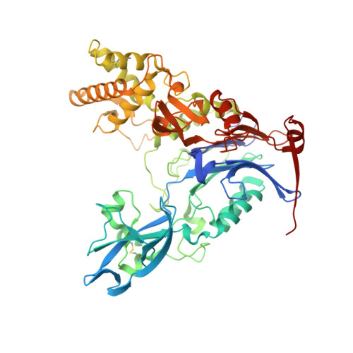
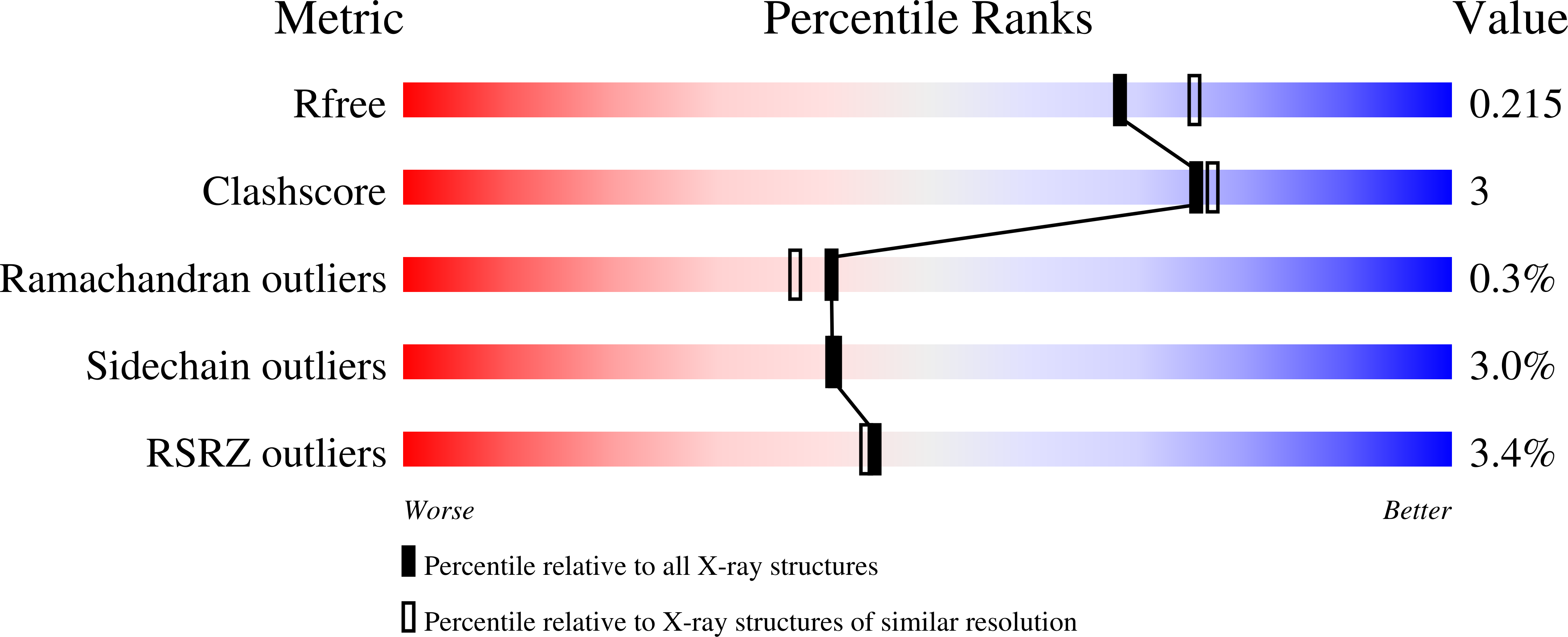
    The enzyme PvdQ (E.C. 3.5.1.97) from Pseudomonas aeruginosa is an N-terminal nucleophile hydrolase that catalyzes the removal of an N-myristyl substituent from a biosynthetic precursor of the iron-chelating siderophore pyoverdine. Inhibitors of pyoverdine biosynthesis are potential antibiotics since iron is essential for growth and scarce in most infections. PvdQ also catalyzes hydrolytic amide bond cleavage of selected N-acyl-l-homoserine lactone quorum-sensing signals used by some Gram-negative pathogens to coordinate the transcription of virulence factors. The resulting quorum-quenching activity of PvdQ has potential applications in antivirulence therapies. To inform both inhibitor design and enzyme engineering efforts, a series of n-alkylboronic acid inhibitors of PvdQ was characterized to reveal determinants of ligand selectivity. A simple homologation series results in compounds with Ki values that span from 4.7 mM to 190 pM, with a dependence of ΔGbind values on chain length of -1.0 kcal/mol/CH2. X-ray crystal structures are determined for the PvdQ complexes with 1-ethyl-, 1-butyl-, 1-hexyl-, and 1-octylboronic acids at 1.6, 1.8, 2.0, and 2.1 Å resolution, respectively. The 1-hexyl- and 1-octylboronic acids form tetrahedral adducts with the active-site N-terminal Ser217 in the β-subunit of PvdQ, and the n-alkyl substituents are bound in the acyl-group binding site. The 1-ethyl- and 1-butylboronic acids also form adducts with Ser217 but instead form trigonal planar adducts and extend their n-alkyl substituents into an alternative binding site. These results are interpreted to propose a ligand discrimination model for PvdQ that informs the development of PvdQ-related tools and therapeutics.


  • Organizational Affiliation

    Medicinal Chemistry Division, College of Pharmacy, †Biochemistry Graduate Program, and ⊥Center for Infectious Disease, The University of Texas , Austin, Texas 78712, United States.


Macromolecules
Find similar proteins by:  (by identity cutoff)  |  3D Structure
Entity ID: 1
MoleculeChains Sequence LengthOrganismDetailsImage
Acyl-homoserine lactone acylase PvdQ165Pseudomonas aeruginosa PAO1Mutation(s): 0 
Gene Names: pvdQqsc112PA2385
EC: 3.5.1.97
UniProt
Find proteins for Q9I194 (Pseudomonas aeruginosa (strain ATCC 15692 / DSM 22644 / CIP 104116 / JCM 14847 / LMG 12228 / 1C / PRS 101 / PAO1))
Explore Q9I194 
Go to UniProtKB:  Q9I194
Entity Groups  
Sequence Clusters30% Identity50% Identity70% Identity90% Identity95% Identity100% Identity
UniProt GroupQ9I194
Sequence Annotations
Expand
  • Reference Sequence
Find similar proteins by:  (by identity cutoff)  |  3D Structure
Entity ID: 2
MoleculeChains Sequence LengthOrganismDetailsImage
Acyl-homoserine lactone acylase PvdQ548Pseudomonas aeruginosa PAO1Mutation(s): 0 
Gene Names: pvdQqsc112PA2385
EC: 3.5.1.97
UniProt
Find proteins for Q9I194 (Pseudomonas aeruginosa (strain ATCC 15692 / DSM 22644 / CIP 104116 / JCM 14847 / LMG 12228 / 1C / PRS 101 / PAO1))
Explore Q9I194 
Go to UniProtKB:  Q9I194
Entity Groups  
Sequence Clusters30% Identity50% Identity70% Identity90% Identity95% Identity100% Identity
UniProt GroupQ9I194
Sequence Annotations
Expand
  • Reference Sequence
Small Molecules
Ligands 2 Unique
IDChains Name / Formula / InChI Key2D Diagram3D Interactions
3QJ
Query on 3QJ

Download Ideal Coordinates CCD File 
G [auth B]hexyl(trihydroxy)borate(1-)
C6 H16 B O3
ZVNFLSCBVLLZCO-UHFFFAOYSA-N
GOL
Query on GOL

Download Ideal Coordinates CCD File 
C [auth A]
D [auth A]
E [auth A]
F [auth A]
H [auth B]
C [auth A],
D [auth A],
E [auth A],
F [auth A],
H [auth B],
I [auth B],
J [auth B],
K [auth B],
L [auth B],
M [auth B],
N [auth B],
O [auth B],
P [auth B],
Q [auth B],
R [auth B],
S [auth B]
GLYCEROL
C3 H8 O3
PEDCQBHIVMGVHV-UHFFFAOYSA-N
Experimental Data & Validation

Experimental Data

  • Method: X-RAY DIFFRACTION
  • Resolution: 2.00 Å
  • R-Value Free: 0.215 
  • R-Value Work: 0.180 
  • R-Value Observed: 0.182 
  • Space Group: C 2 2 21
Unit Cell:
Length ( Å )Angle ( ˚ )
a = 121.131α = 90
b = 168.243β = 90
c = 94.945γ = 90
Software Package:
Software NamePurpose
PHENIXrefinement

Structure Validation

View Full Validation Report


Entry History 

Deposition Data

Revision History  (Full details and data files)

  • Version 1.0: 2014-11-12
    Type: Initial release
  • Version 1.1: 2023-12-27
    Changes: Data collection, Database references, Derived calculations, Other, Refinement description, Source and taxonomy, Structure summary