4WK3

Structure of Staphyloccus aureus PstA


Experimental Data Snapshot

  • Method: X-RAY DIFFRACTION
  • Resolution: 2.60 Å
  • R-Value Free: 0.292 
  • R-Value Work: 0.252 
  • R-Value Observed: 0.256 

wwPDB Validation   3D Report Full Report


This is version 2.0 of the entry. See complete history


Literature

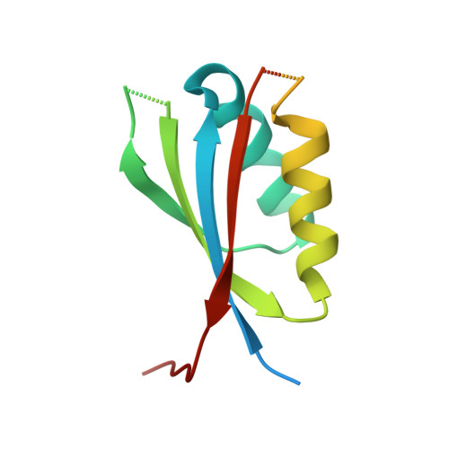
c-di-AMP recognition by Staphylococcus aureus PstA.

Muller, M.Hopfner, K.Witte, G.

(2015) FEBS Lett 589: 45-51

  • DOI: https://doi.org/10.1016/j.febslet.2014.11.022
  • Primary Citation of Related Structures:  
    4WK1, 4WK3

  • PubMed Abstract: 

    Cyclic-di-AMP (c-di-AMP) is a bacterial secondary messenger involved in various processes, including sensing of DNA-integrity, cell wall metabolism and potassium transport. A number of c-di-AMP receptor proteins have recently been identified in Staphylococcus aureus. One of them - PstA - possesses a ferredoxin-like fold and is structurally related to the class of PII signal-transduction proteins. PII proteins are involved in a large number of pathways, most of them associated with nitrogen metabolism. In this study we describe the mode of c-di-AMP binding and subsequent structural changes of S. aureus PstA. An altered architecture in PstA compared to canonical PII proteins results in differences in ligand coordination.


  • Organizational Affiliation

    Ludwig-Maximilians-Universität München, Gene Center and Dept. of Biochemistry, Feodor-Lynen-Str. 25, 81377 Munich, Germany.


Macromolecules
Find similar proteins by:  (by identity cutoff)  |  3D Structure
Entity ID: 1
MoleculeChains Sequence LengthOrganismDetailsImage
PstA129Staphylococcus aureus subsp. aureus COLMutation(s): 0 
UniProt
Find proteins for A0A0H2WXZ7 (Staphylococcus aureus (strain COL))
Explore A0A0H2WXZ7 
Go to UniProtKB:  A0A0H2WXZ7
Entity Groups  
Sequence Clusters30% Identity50% Identity70% Identity90% Identity95% Identity100% Identity
UniProt GroupA0A0H2WXZ7
Sequence Annotations
Expand
  • Reference Sequence
Small Molecules
Ligands 1 Unique
IDChains Name / Formula / InChI Key2D Diagram3D Interactions
CL
Query on CL

Download Ideal Coordinates CCD File 
B [auth A]CHLORIDE ION
Cl
VEXZGXHMUGYJMC-UHFFFAOYSA-M
Experimental Data & Validation

Experimental Data

  • Method: X-RAY DIFFRACTION
  • Resolution: 2.60 Å
  • R-Value Free: 0.292 
  • R-Value Work: 0.252 
  • R-Value Observed: 0.256 
  • Space Group: P 43 3 2
Unit Cell:
Length ( Å )Angle ( ˚ )
a = 99.32α = 90
b = 99.32β = 90
c = 99.32γ = 90
Software Package:
Software NamePurpose
PHENIXrefinement
XDSdata reduction
XSCALEdata scaling
PHASERphasing
Cootmodel building

Structure Validation

View Full Validation Report


Entry History & Funding Information

Deposition Data


Funding OrganizationLocationGrant Number
German Research FoundationGermany3717/2-1
German Research FoundationGermanyGRK1721

Revision History  (Full details and data files)

  • Version 1.0: 2014-11-26
    Type: Initial release
  • Version 1.1: 2014-12-24
    Changes: Database references
  • Version 1.2: 2014-12-31
    Changes: Database references
  • Version 2.0: 2024-01-10
    Changes: Atomic model, Author supporting evidence, Data collection, Database references, Refinement description