


Structural and Functional Characterization of Acinetobacter baumannii Nucleoside Diphosphate Kinase
Hu, Y., Feng, F., Liu, Y.(2015) PROG BIOCHEM BIOPHYS 42: 260-267
Experimental Data Snapshot
wwPDB Validation 3D Report Full Report

Entity ID: 1 | |||||
|---|---|---|---|---|---|
| Molecule | Chains | Sequence Length | Organism | Details | Image |
| Nucleoside diphosphate kinase | 142 | Acinetobacter baumannii SDF | Mutation(s): 0 Gene Names: ndk, ABSDF3006 EC: 2.7.4.6 | 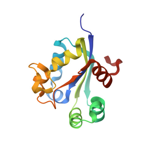 | |
UniProt | |||||
Find proteins for B0VKS3 (Acinetobacter baumannii (strain SDF)) Explore B0VKS3 Go to UniProtKB: B0VKS3 | |||||
Entity Groups | |||||
| Sequence Clusters | 30% Identity50% Identity70% Identity90% Identity95% Identity100% Identity | ||||
| UniProt Group | B0VKS3 | ||||
Sequence AnnotationsExpand | |||||
| |||||
| Length ( Å ) | Angle ( ˚ ) |
|---|---|
| a = 60.33 | α = 90 |
| b = 75.4 | β = 90 |
| c = 82.12 | γ = 90 |
| Software Name | Purpose |
|---|---|
| PHENIX | refinement |
RCSB PDB (citation) is hosted by
RCSB PDB is a member of the

