4WBC

2.13 A STRUCTURE OF A KUNITZ-TYPE WINGED BEAN CHYMOTRYPSIN INHIBITOR PROTEIN


Experimental Data Snapshot

  • Method: X-RAY DIFFRACTION
  • Resolution: 2.14 Å
  • R-Value Free: 0.257 
  • R-Value Work: 0.199 

wwPDB Validation   3D Report Full Report


This is version 1.5 of the entry. See complete history


Literature

Cryocrystallography of a Kunitz-type serine protease inhibitor: the 90 K structure of winged bean chymotrypsin inhibitor (WCI) at 2.13 A resolution.

Ravichandran, S.Sen, U.Chakrabarti, C.Dattagupta, J.K.

(1999) Acta Crystallogr D Biol Crystallogr 55: 1814-1821

  • DOI: https://doi.org/10.1107/s0907444999009877
  • Primary Citation of Related Structures:  
    4WBC

  • PubMed Abstract: 

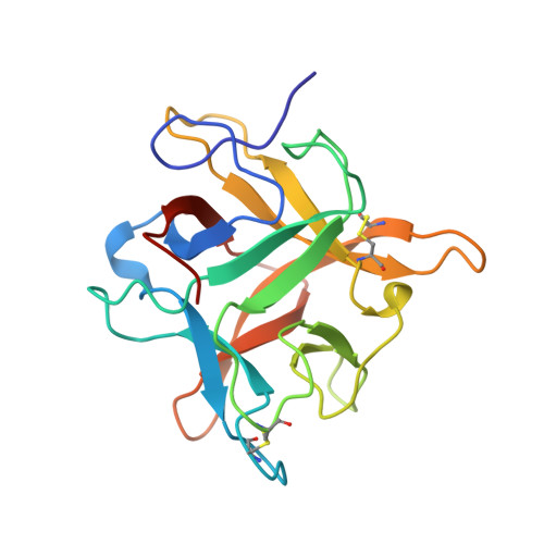
    The crystal structure of a Kunitz-type double-headed alpha--chymotrypsin inhibitor from winged bean seeds has been refined at 2.13 A resolution using data collected from cryo-cooled (90 K) crystals which belong to the hexagonal space group P6(1)22 with unit-cell parameters a = b = 60.84, c = 207.91 A. The volume of the unit cell is reduced by 5.3% on cooling. The refinement converged to an R value of 20.0% (R(free) = 25.8%) for 11100 unique reflections and the model shows good stereochemistry, with r.m.s. deviations from ideal values for bond lengths and bond angles of 0.011 A and 1.4 degrees, respectively. The structural architecture of the protein consists of 12 antiparallel beta-strands joined in the form of a characteristic beta-trefoil fold, with the two reactive-site regions, Asn38-Leu43 and Gln63-Phe68, situated on two external loops. Although the overall protein fold is the same as that of the room-temperature model, some conformational changes are observed in the loop regions and in the side chains of a few surface residues. A total of 176 ordered water molecules and five sulfate ions are included in the model.


  • Organizational Affiliation

    Crystallography Division, Saha Institute of Nuclear Physics, 1/AF Bidhan Nagar, Calcutta 700 064, India.


Macromolecules
Find similar proteins by:  (by identity cutoff)  |  3D Structure
Entity ID: 1
MoleculeChains Sequence LengthOrganismDetailsImage
PROTEIN (CHYMOTRYPSIN INHIBITOR)183Psophocarpus tetragonolobusMutation(s): 0 
UniProt
Find proteins for P10822 (Psophocarpus tetragonolobus)
Explore P10822 
Go to UniProtKB:  P10822
Entity Groups  
Sequence Clusters30% Identity50% Identity70% Identity90% Identity95% Identity100% Identity
UniProt GroupP10822
Sequence Annotations
Expand
  • Reference Sequence
Experimental Data & Validation

Experimental Data

  • Method: X-RAY DIFFRACTION
  • Resolution: 2.14 Å
  • R-Value Free: 0.257 
  • R-Value Work: 0.199 
  • Space Group: P 61 2 2
Unit Cell:
Length ( Å )Angle ( ˚ )
a = 60.84α = 90
b = 60.84β = 90
c = 207.91γ = 120
Software Package:
Software NamePurpose
DENZOdata reduction
SCALEPACKdata scaling
REFMACrefinement

Structure Validation

View Full Validation Report


Entry History 

Deposition Data

Revision History  (Full details and data files)

  • Version 1.0: 1999-03-12
    Type: Initial release
  • Version 1.1: 2007-10-16
    Changes: Version format compliance
  • Version 1.2: 2011-07-13
    Changes: Derived calculations, Version format compliance
  • Version 1.3: 2018-04-04
    Changes: Data collection
  • Version 1.4: 2019-11-06
    Changes: Data collection, Database references
  • Version 1.5: 2023-12-27
    Changes: Data collection, Database references, Derived calculations