4WBB

Single Turnover Autophosphorylation Cycle of the PKA RIIb Holoenzyme


Experimental Data Snapshot

  • Method: X-RAY DIFFRACTION
  • Resolution: 2.80 Å
  • R-Value Free: 0.279 
  • R-Value Work: 0.232 

wwPDB Validation   3D Report Full Report


Ligand Structure Quality Assessment 


This is version 1.5 of the entry. See complete history


Literature

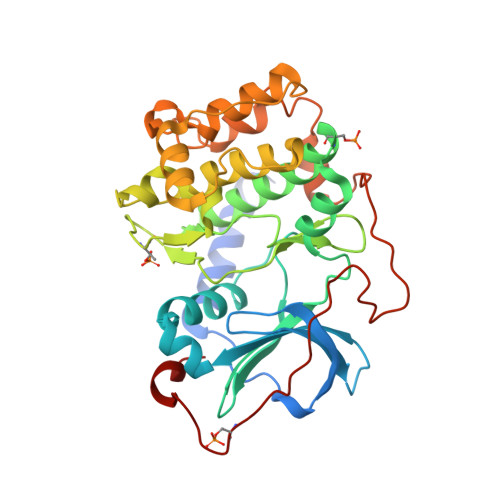
Single Turnover Autophosphorylation Cycle of the PKA RII beta Holoenzyme.

Zhang, P.Knape, M.J.Ahuja, L.G.Keshwani, M.M.King, C.C.Sastri, M.Herberg, F.W.Taylor, S.S.

(2015) PLoS Biol 13: e1002192-e1002192

  • DOI: https://doi.org/10.1371/journal.pbio.1002192
  • Primary Citation of Related Structures:  
    4WBB

  • PubMed Abstract: 

    To provide tight spatiotemporal signaling control, the cyclic adenosine monophosphate (cAMP)-dependent protein kinase (PKA) holoenzyme typically nucleates a macromolecular complex or a "PKA signalosome." Using the RIIβ holoenzyme as a prototype, we show how autophosphorylation/dephosphorylation of the RIIβ subunit, as well as cAMP and metal ions, contribute to the dynamics of PKA signaling. While we showed previously that the RIIβ holoenzyme could undergo a single turnover autophosphorylation with adenosine triphosphate and magnesium (MgATP) and trap both products in the crystal lattice, we asked here whether calcium could trap an ATP:RIIβ holoenzyme since the RIIβ holoenzyme is located close to ion channels. The 2.8Å structure of an RIIβp2:C2:(Ca2ADP)2 holoenzyme, supported by biochemical and biophysical data, reveals a trapped single phosphorylation event similar to MgATP. Thus, calcium can mediate a single turnover event with either ATP or adenosine-5'-(β,γ-imido)triphosphate (AMP-PNP), even though it cannot support steady-state catalysis efficiently. The holoenzyme serves as a "product trap" because of the slow off-rate of the pRIIβ subunit, which is controlled by cAMP, not by phosphorylation of the inhibitor site. By quantitatively defining the RIIβ signaling cycle, we show that release of pRIIβ in the presence of cAMP is reduced by calcium, whereas autophosphorylation at the phosphorylation site (P-site) inhibits holoenzyme reassociation with the catalytic subunit. Adding a single phosphoryl group to the preformed RIIβ holoenzyme thus creates a signaling cycle in which phosphatases become an essential partner. This previously unappreciated molecular mechanism is an integral part of PKA signaling for type II holoenzymes.


  • Organizational Affiliation

    Department of Pharmacology, University of California at San Diego, La Jolla, California, United States of America.


Macromolecules
Find similar proteins by:  (by identity cutoff)  |  3D Structure
Entity ID: 1
MoleculeChains Sequence LengthOrganismDetailsImage
cAMP-dependent protein kinase type II-beta regulatory subunit416Mus musculusMutation(s): 1 
Gene Names: Prkar2b
UniProt
Find proteins for P31324 (Mus musculus)
Explore P31324 
Go to UniProtKB:  P31324
Entity Groups  
Sequence Clusters30% Identity50% Identity70% Identity90% Identity95% Identity100% Identity
UniProt GroupP31324
Sequence Annotations
Expand
  • Reference Sequence
Find similar proteins by:  (by identity cutoff)  |  3D Structure
Entity ID: 2
MoleculeChains Sequence LengthOrganismDetailsImage
cAMP-dependent protein kinase catalytic subunit alpha350Mus musculusMutation(s): 0 
Gene Names: PrkacaPkaca
EC: 2.7.11.11
UniProt
Find proteins for P05132 (Mus musculus)
Explore P05132 
Go to UniProtKB:  P05132
Entity Groups  
Sequence Clusters30% Identity50% Identity70% Identity90% Identity95% Identity100% Identity
UniProt GroupP05132
Sequence Annotations
Expand
  • Reference Sequence
Small Molecules
Modified Residues  2 Unique
IDChains TypeFormula2D DiagramParent
SEP
Query on SEP
A
L-PEPTIDE LINKINGC3 H8 N O6 PSER
TPO
Query on TPO
B
L-PEPTIDE LINKINGC4 H10 N O6 PTHR
Experimental Data & Validation

Experimental Data

  • Method: X-RAY DIFFRACTION
  • Resolution: 2.80 Å
  • R-Value Free: 0.279 
  • R-Value Work: 0.232 
  • Space Group: C 2 2 2
Unit Cell:
Length ( Å )Angle ( ˚ )
a = 150.38α = 90
b = 213.951β = 90
c = 61.967γ = 90
Software Package:
Software NamePurpose
PHASERphasing

Structure Validation

View Full Validation Report


Ligand Structure Quality Assessment 


Entry History & Funding Information

Deposition Data


Funding OrganizationLocationGrant Number
National Institutes of Health/National Institute of General Medical Sciences (NIH/NIGMS)United StatesGM034921

Revision History  (Full details and data files)

  • Version 1.0: 2015-05-20
    Type: Initial release
  • Version 1.1: 2015-07-22
    Changes: Database references
  • Version 1.2: 2015-08-26
    Changes: Experimental preparation
  • Version 1.3: 2017-09-20
    Changes: Author supporting evidence, Derived calculations, Refinement description
  • Version 1.4: 2019-12-25
    Changes: Author supporting evidence
  • Version 1.5: 2023-12-27
    Changes: Data collection, Database references, Derived calculations