4V3O

Designed armadillo repeat protein with 5 internal repeats, 2nd generation C-cap and 3rd generation N-cap.


Experimental Data Snapshot

  • Method: X-RAY DIFFRACTION
  • Resolution: 2.00 Å
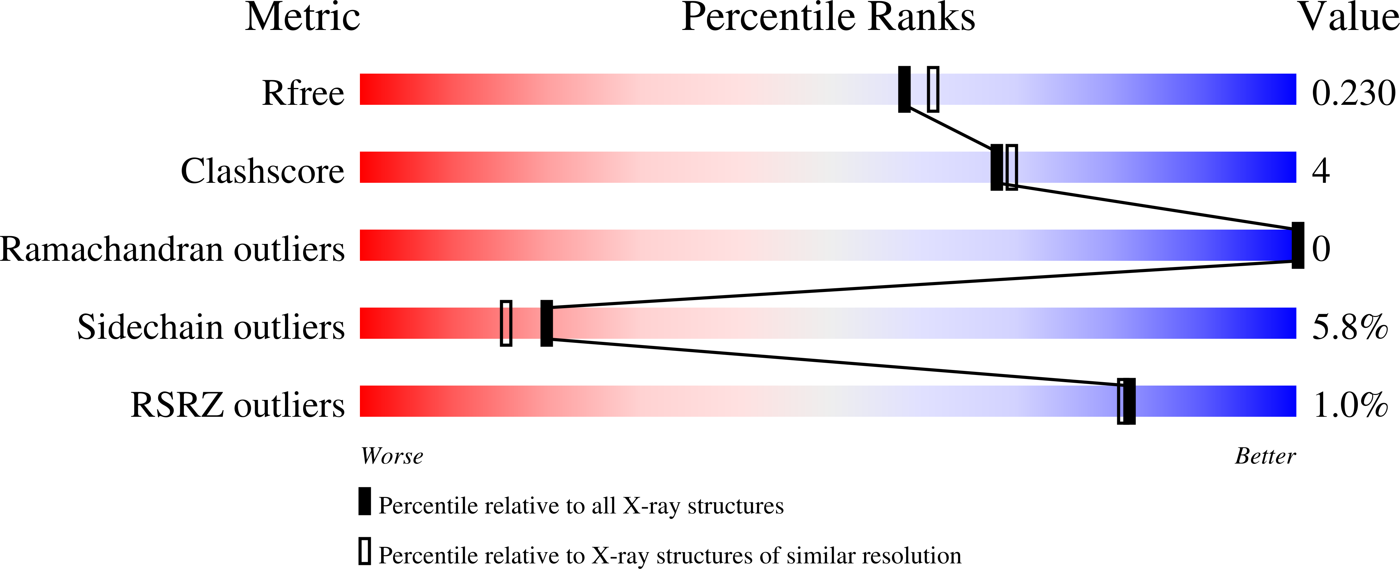
  • R-Value Free: 0.224 
  • R-Value Work: 0.168 
  • R-Value Observed: 0.171 

wwPDB Validation   3D Report Full Report


This is version 1.2 of the entry. See complete history


Literature

Structures of Designed Armadillo-Repeat Proteins Show Propagation of Inter-Repeat Interface Effects

Reichen, C.Madhurantakam, C.Hansen, S.Grutter, M.G.Pluckthun, A.Mittl, P.R.E.

(2016) Acta Crystallogr D Biol Crystallogr 72: 168

  • DOI: https://doi.org/10.1107/S2059798315023116
  • Primary Citation of Related Structures:  
    4V3O, 4V3Q, 4V3R

  • PubMed Abstract: 

    The armadillo repeat serves as a scaffold for the development of modular peptide-recognition modules. In order to develop such a system, three crystal structures of designed armadillo-repeat proteins with third-generation N-caps (YIII-type), four or five internal repeats (M-type) and second-generation C-caps (AII-type) were determined at 1.8 Å (His-YIIIM4AII), 2.0 Å (His-YIIIM5AII) and 1.95 Å (YIIIM5AII) resolution and compared with those of variants with third-generation C-caps. All constructs are full consensus designs in which the internal repeats have exactly the same sequence, and hence identical conformations of the internal repeats are expected. The N-cap and internal repeats M1 to M3 are indeed extremely similar, but the comparison reveals structural differences in internal repeats M4 and M5 and the C-cap. These differences are caused by long-range effects of the C-cap, contacting molecules in the crystal, and the intrinsic design of the repeat. Unfortunately, the rigid-body movement of the C-terminal part impairs the regular arrangement of internal repeats that forms the putative peptide-binding site. The second-generation C-cap improves the packing of buried residues and thereby the stability of the protein. These considerations are useful for future improvements of an armadillo-repeat-based peptide-recognition system.


  • Organizational Affiliation

    Department of Biochemistry, University of Zürich, Winterthurerstrasse 190, 8057 Zürich, Switzerland.


Macromolecules
Find similar proteins by:  (by identity cutoff)  |  3D Structure
Entity ID: 1
MoleculeChains Sequence LengthOrganismDetailsImage
YIII_M5_AII
A, B, C, D
289synthetic constructMutation(s): 0 
Entity Groups  
Sequence Clusters30% Identity50% Identity70% Identity90% Identity95% Identity100% Identity
Sequence Annotations
Expand
  • Reference Sequence
Small Molecules
Ligands 2 Unique
IDChains Name / Formula / InChI Key2D Diagram3D Interactions
ACT
Query on ACT

Download Ideal Coordinates CCD File 
Q [auth D]ACETATE ION
C2 H3 O2
QTBSBXVTEAMEQO-UHFFFAOYSA-M
CA
Query on CA

Download Ideal Coordinates CCD File 
E [auth A]
F [auth A]
G [auth A]
H [auth A]
I [auth B]
E [auth A],
F [auth A],
G [auth A],
H [auth A],
I [auth B],
J [auth B],
K [auth B],
L [auth B],
M [auth B],
N [auth C],
O [auth C],
P [auth D]
CALCIUM ION
Ca
BHPQYMZQTOCNFJ-UHFFFAOYSA-N
Experimental Data & Validation

Experimental Data

  • Method: X-RAY DIFFRACTION
  • Resolution: 2.00 Å
  • R-Value Free: 0.224 
  • R-Value Work: 0.168 
  • R-Value Observed: 0.171 
  • Space Group: P 41
Unit Cell:
Length ( Å )Angle ( ˚ )
a = 102.586α = 90
b = 102.586β = 90
c = 111.107γ = 90
Software Package:
Software NamePurpose
REFMACrefinement
MOSFLMdata reduction
SCALAdata scaling
PHENIXphasing

Structure Validation

View Full Validation Report


Entry History 

Deposition Data

Revision History  (Full details and data files)

  • Version 1.0: 2016-01-13
    Type: Initial release
  • Version 1.1: 2016-03-02
    Changes: Database references
  • Version 1.2: 2024-01-10
    Changes: Data collection, Database references, Derived calculations, Other, Refinement description