4UZI

Crystal Structure of AauDyP Complexed with Imidazole


Experimental Data Snapshot

  • Method: X-RAY DIFFRACTION
  • Resolution: 2.10 Å
  • R-Value Free: 0.239 
  • R-Value Work: 0.179 
  • R-Value Observed: 0.182 

wwPDB Validation   3D Report Full Report


Ligand Structure Quality Assessment 


This is version 3.1 of the entry. See complete history


Literature

The Toolbox of Auricularia Auricula-Judae Dye-Decolorizing Peroxidase - Identification of Three New Potential Substrate-Interaction Sites.

Strittmatter, E.Serrer, K.Liers, C.Ullrich, R.Hofrichter, M.Piontek, K.Schleicher, E.Plattner, D.A.

(2015) Arch Biochem Biophys 574: 75

  • DOI: https://doi.org/10.1016/j.abb.2014.12.016
  • Primary Citation of Related Structures:  
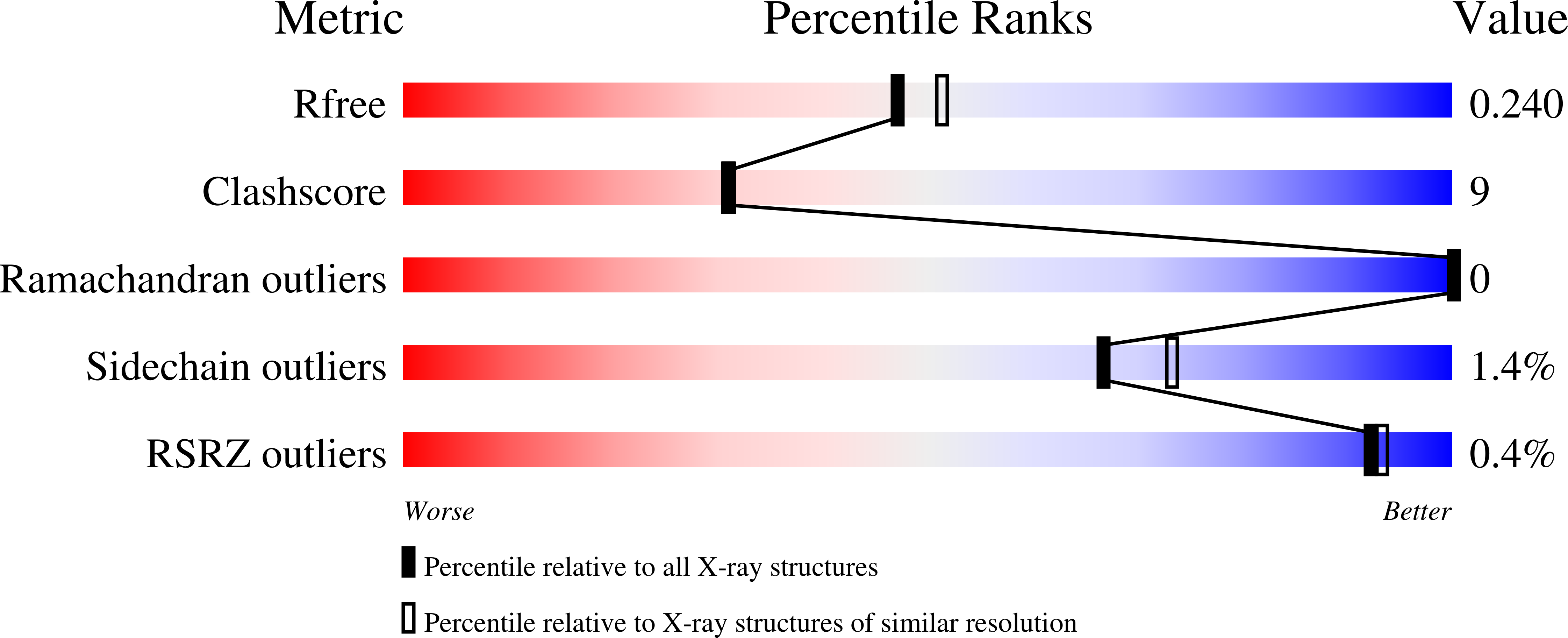
    4UZI

  • PubMed Abstract: 

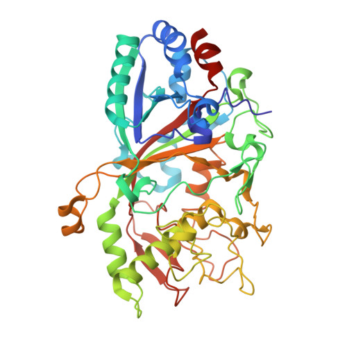
    Dye-decolorizing peroxidases (DyPs) such as AauDyPI from the fungus Auricularia auricula-judae are able to oxidize substrates of different kinds and sizes. A crystal structure of an AauDyPI-imidazole complex gives insight into the binding patterns of organic molecules within the heme cavity of a DyP. Several small N-containing heterocyclic aromatics are shown to bind in the AauDyPI heme cavity, hinting to susceptibility of DyPs to azole-based inhibitors similar to cytochromes P450. Imidazole is confirmed as a competitive inhibitor with regard to peroxide binding. In contrast, bulky substrates such as anthraquinone dyes are converted at the enzyme surface. In the crystal structure a substrate analog, 4-(2-hydroxyethyl)-1-piperazineethanesulfonic acid (HEPES), binds to a tyrosine-rich hollow harboring Y25, Y147, and Y337. Spin trapping with a nitric oxide donor uncovers Y229 as an additional tyrosine-based radical center in AauDyPI. Multi-frequency EPR spectroscopy further reveals the presence of at least one intermediate tryptophanyl radical center in activated AauDyPI with W377 as the most likely candidate.


  • Organizational Affiliation

    Institute of Organic Chemistry, University of Freiburg, Albertstrasse 21, 79104 Freiburg, Germany.


Macromolecules
Find similar proteins by:  (by identity cutoff)  |  3D Structure
Entity ID: 1
MoleculeChains Sequence LengthOrganismDetailsImage
DYE-DECOLORIZING PEROXIDASE
A, B
446Auricularia auricula-judaeMutation(s): 0 
EC: 1.11.1.19
UniProt
Find proteins for I2DBY1 (Auricularia auricula-judae)
Explore I2DBY1 
Go to UniProtKB:  I2DBY1
Entity Groups  
Sequence Clusters30% Identity50% Identity70% Identity90% Identity95% Identity100% Identity
UniProt GroupI2DBY1
Sequence Annotations
Expand
  • Reference Sequence
Oligosaccharides

Help

Entity ID: 2
MoleculeChains Length2D Diagram Glycosylation3D Interactions
2-acetamido-2-deoxy-beta-D-glucopyranose-(1-4)-2-acetamido-2-deoxy-beta-D-glucopyranose
C
2N-Glycosylation
Glycosylation Resources
GlyTouCan:  G42666HT
GlyCosmos:  G42666HT
GlyGen:  G42666HT
Entity ID: 3
MoleculeChains Length2D Diagram Glycosylation3D Interactions
beta-D-mannopyranose-(1-4)-2-acetamido-2-deoxy-beta-D-glucopyranose-(1-4)-2-acetamido-2-deoxy-beta-D-glucopyranose
D
3N-Glycosylation
Glycosylation Resources
GlyTouCan:  G15407YE
GlyCosmos:  G15407YE
GlyGen:  G15407YE
Small Molecules
Ligands 11 Unique
IDChains Name / Formula / InChI Key2D Diagram3D Interactions
HEM
Query on HEM

Download Ideal Coordinates CCD File 
E [auth A],
T [auth B]
PROTOPORPHYRIN IX CONTAINING FE
C34 H32 Fe N4 O4
KABFMIBPWCXCRK-RGGAHWMASA-L
EPE
Query on EPE

Download Ideal Coordinates CCD File 
DA [auth B],
N [auth A]
4-(2-HYDROXYETHYL)-1-PIPERAZINE ETHANESULFONIC ACID
C8 H18 N2 O4 S
JKMHFZQWWAIEOD-UHFFFAOYSA-N
NAG
Query on NAG

Download Ideal Coordinates CCD File 
F [auth A],
G [auth A],
U [auth B],
V [auth B]
2-acetamido-2-deoxy-beta-D-glucopyranose
C8 H15 N O6
OVRNDRQMDRJTHS-FMDGEEDCSA-N
MRD
Query on MRD

Download Ideal Coordinates CCD File 
J [auth A](4R)-2-METHYLPENTANE-2,4-DIOL
C6 H14 O2
SVTBMSDMJJWYQN-RXMQYKEDSA-N
MPD
Query on MPD

Download Ideal Coordinates CCD File 
AA [auth B]
BA [auth B]
CA [auth B]
K [auth A]
L [auth A]
AA [auth B],
BA [auth B],
CA [auth B],
K [auth A],
L [auth A],
Y [auth B],
Z [auth B]
(4S)-2-METHYL-2,4-PENTANEDIOL
C6 H14 O2
SVTBMSDMJJWYQN-YFKPBYRVSA-N
OXD
Query on OXD

Download Ideal Coordinates CCD File 
R [auth A]OXALIC ACID
C2 H2 O4
MUBZPKHOEPUJKR-UHFFFAOYSA-N
GOA
Query on GOA

Download Ideal Coordinates CCD File 
HA [auth B]GLYCOLIC ACID
C2 H4 O3
AEMRFAOFKBGASW-UHFFFAOYSA-N
IMD
Query on IMD

Download Ideal Coordinates CCD File 
H [auth A],
I [auth A],
W [auth B],
X [auth B]
IMIDAZOLE
C3 H5 N2
RAXXELZNTBOGNW-UHFFFAOYSA-O
ACT
Query on ACT

Download Ideal Coordinates CCD File 
EA [auth B]
FA [auth B]
IA [auth B]
O [auth A]
P [auth A]
EA [auth B],
FA [auth B],
IA [auth B],
O [auth A],
P [auth A],
Q [auth A]
ACETATE ION
C2 H3 O2
QTBSBXVTEAMEQO-UHFFFAOYSA-M
FMT
Query on FMT

Download Ideal Coordinates CCD File 
M [auth A]FORMIC ACID
C H2 O2
BDAGIHXWWSANSR-UHFFFAOYSA-N
K
Query on K

Download Ideal Coordinates CCD File 
GA [auth B],
S [auth A]
POTASSIUM ION
K
NPYPAHLBTDXSSS-UHFFFAOYSA-N
Binding Affinity Annotations 
IDSourceBinding Affinity
IMD Binding MOAD:  4UZI Ki: 5.78e+6 (nM) from 1 assay(s)
Experimental Data & Validation

Experimental Data

  • Method: X-RAY DIFFRACTION
  • Resolution: 2.10 Å
  • R-Value Free: 0.239 
  • R-Value Work: 0.179 
  • R-Value Observed: 0.182 
  • Space Group: P 1 21 1
Unit Cell:
Length ( Å )Angle ( ˚ )
a = 67.33α = 90
b = 45.71β = 91.61
c = 140.47γ = 90
Software Package:
Software NamePurpose
REFMACrefinement
XDSdata reduction
XDSdata scaling
PHASERphasing

Structure Validation

View Full Validation Report


Ligand Structure Quality Assessment 


Entry History 

Deposition Data

Revision History  (Full details and data files)

  • Version 1.0: 2015-01-14
    Type: Initial release
  • Version 1.1: 2015-05-13
    Changes: Database references
  • Version 2.0: 2020-07-29
    Type: Remediation
    Reason: Carbohydrate remediation
    Changes: Advisory, Atomic model, Data collection, Derived calculations, Other, Structure summary
  • Version 3.0: 2023-11-15
    Changes: Atomic model, Data collection, Database references, Structure summary
  • Version 3.1: 2024-01-10
    Changes: Refinement description