4UU5

CRYSTAL STRUCTURE OF THE PDZ DOMAIN OF PALS1 IN COMPLEX WITH THE CRB PEPTIDE


Experimental Data Snapshot

  • Method: X-RAY DIFFRACTION
  • Resolution: 1.23 Å
  • R-Value Free: 0.183 
  • R-Value Work: 0.168 
  • R-Value Observed: 0.169 

wwPDB Validation   3D Report Full Report


This is version 2.1 of the entry. See complete history


Literature

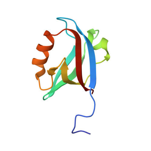
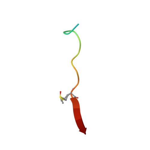
Structures of the human Pals1 PDZ domain with and without ligand suggest gated access of Crb to the PDZ peptide-binding groove.

Ivanova, M.E.Fletcher, G.C.O'Reilly, N.Purkiss, A.G.Thompson, B.J.McDonald, N.Q.

(2015) Acta Crystallogr D Biol Crystallogr 71: 555-564

  • DOI: https://doi.org/10.1107/S139900471402776X
  • Primary Citation of Related Structures:  
    4UU5, 4UU6

  • PubMed Abstract: 

    Many components of epithelial polarity protein complexes possess PDZ domains that are required for protein interaction and recruitment to the apical plasma membrane. Apical localization of the Crumbs (Crb) transmembrane protein requires a PDZ-mediated interaction with Pals1 (protein-associated with Lin7, Stardust, MPP5), a member of the p55 family of membrane-associated guanylate kinases (MAGUKs). This study describes the molecular interaction between the Crb carboxy-terminal motif (ERLI), which is required for Drosophila cell polarity, and the Pals1 PDZ domain using crystallography and fluorescence polarization. Only the last four Crb residues contribute to Pals1 PDZ-domain binding affinity, with specificity contributed by conserved charged interactions. Comparison of the Crb-bound Pals1 PDZ structure with an apo Pals1 structure reveals a key Phe side chain that gates access to the PDZ peptide-binding groove. Removal of this side chain enhances the binding affinity by more than fivefold, suggesting that access of Crb to Pals1 may be regulated by intradomain contacts or by protein-protein interaction.


  • Organizational Affiliation

    Structural Biology Laboratories, Cancer Research UK, 44 Lincoln's Inn Fields, London WC2A 3LY, England.


Macromolecules
Find similar proteins by:  (by identity cutoff)  |  3D Structure
Entity ID: 1
MoleculeChains Sequence LengthOrganismDetailsImage
MAGUK P55 SUBFAMILY MEMBER 590Homo sapiensMutation(s): 0 
UniProt & NIH Common Fund Data Resources
Find proteins for Q8N3R9 (Homo sapiens)
Explore Q8N3R9 
Go to UniProtKB:  Q8N3R9
PHAROS:  Q8N3R9
GTEx:  ENSG00000072415 
Entity Groups  
Sequence Clusters30% Identity50% Identity70% Identity90% Identity95% Identity100% Identity
UniProt GroupQ8N3R9
Sequence Annotations
Expand
  • Reference Sequence

Find similar proteins by:  Sequence   |   3D Structure  

Entity ID: 2
MoleculeChains Sequence LengthOrganismDetailsImage
PROTEIN CRUMBS HOMOLOG 117Homo sapiensMutation(s): 0 
UniProt & NIH Common Fund Data Resources
Find proteins for P82279 (Homo sapiens)
Explore P82279 
Go to UniProtKB:  P82279
PHAROS:  P82279
GTEx:  ENSG00000134376 
Entity Groups  
Sequence Clusters30% Identity50% Identity70% Identity90% Identity95% Identity100% Identity
UniProt GroupP82279
Sequence Annotations
Expand
  • Reference Sequence
Small Molecules
Ligands 3 Unique
IDChains Name / Formula / InChI Key2D Diagram3D Interactions
PUN
Query on PUN

Download Ideal Coordinates CCD File 
K [auth A]2,2,4,4,6,6,8-heptamethylnonane
C16 H34
RDUBCTHNBWFPTP-UHFFFAOYSA-N
CL
Query on CL

Download Ideal Coordinates CCD File 
C [auth A]
D [auth A]
E [auth A]
F [auth A]
G [auth A]
C [auth A],
D [auth A],
E [auth A],
F [auth A],
G [auth A],
H [auth A],
I [auth A],
L [auth B]
CHLORIDE ION
Cl
VEXZGXHMUGYJMC-UHFFFAOYSA-M
NA
Query on NA

Download Ideal Coordinates CCD File 
J [auth A]SODIUM ION
Na
FKNQFGJONOIPTF-UHFFFAOYSA-N
Modified Residues  1 Unique
IDChains TypeFormula2D DiagramParent
SME
Query on SME
B
L-PEPTIDE LINKINGC5 H11 N O3 SMET
Experimental Data & Validation

Experimental Data

  • Method: X-RAY DIFFRACTION
  • Resolution: 1.23 Å
  • R-Value Free: 0.183 
  • R-Value Work: 0.168 
  • R-Value Observed: 0.169 
  • Space Group: P 41 2 2
Unit Cell:
Length ( Å )Angle ( ˚ )
a = 74.73α = 90
b = 74.73β = 90
c = 42.71γ = 90
Software Package:
Software NamePurpose
PHENIXrefinement
XDSdata reduction
XSCALEdata scaling
PHASERphasing

Structure Validation

View Full Validation Report


Entry History 

Deposition Data

Revision History  (Full details and data files)

  • Version 1.0: 2015-01-14
    Type: Initial release
  • Version 1.1: 2015-03-11
    Changes: Database references
  • Version 1.2: 2015-03-25
    Changes: Database references
  • Version 2.0: 2018-02-28
    Changes: Atomic model, Database references
  • Version 2.1: 2024-01-10
    Changes: Data collection, Database references, Derived calculations, Other, Refinement description