4UP1

Crystal structure of native human Thymidylate synthase in active form


Experimental Data Snapshot

  • Method: X-RAY DIFFRACTION
  • Resolution: 2.99 Å
  • R-Value Free: 0.244 
  • R-Value Work: 0.214 
  • R-Value Observed: 0.216 

wwPDB Validation   3D Report Full Report


This is version 1.3 of the entry. See complete history


Literature

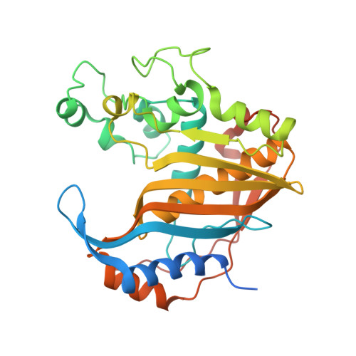
Crystal structure of the active form of native human thymidylate synthase in the absence of bound substrates.

Deschamps, P.Rety, S.Bareille, J.Leulliot, N.

(2017) Acta Crystallogr F Struct Biol Commun 73: 336-341

  • DOI: https://doi.org/10.1107/S2053230X17007233
  • Primary Citation of Related Structures:  
    4UP1

  • PubMed Abstract: 

    Human thymidylate synthase (hTS) provides the sole de novo intracellular source of thymidine 5'-monophosphate (dTMP). hTS is required for DNA replication prior to cell division, making it an attractive target for anticancer chemotherapy and drug discovery. hTS binds 2'-deoxyuridine 5'-monophosphate (dUMP) and the folate co-substrate N 5 ,N 10 -methylenetetrahydrofolate (meTHF) in a pocket near the catalytic residue Cys195. The catalytic loop, which is composed of amino-acid residues 181-197, can adopt two distinct conformations related by a 180° rotation. In the active conformation Cys195 is close to the active site, while in the inactive conformation it is rotated and Cys195 is too distant from the active site for catalysis. Several hTS structures, either native or engineered, have been solved in the active conformation in complex with ligands or inhibitors and at different salt concentrations. However, apo hTS structures have been solved in an inactive conformation in high-salt and low-salt conditions (PDB entries 1ypv, 4h1i, 4gyh, 3egy and 3ehi). Here, the structure of apo hTS crystallized in the active form with sulfate ions coordinated by the arginine residue that binds dUMP is reported.


  • Organizational Affiliation

    Laboratoire de Cristallographie et RMN Biologiques, UMR CNRS 8015, Université Paris Descartes, Sorbonne Paris Cité, Faculté de Pharmacie de Paris, Paris, France.


Macromolecules
Find similar proteins by:  (by identity cutoff)  |  3D Structure
Entity ID: 1
MoleculeChains Sequence LengthOrganismDetailsImage
THYMIDYLATE SYNTHASE
A, B, C, D
313Homo sapiensMutation(s): 1 
EC: 2.1.1.45
UniProt & NIH Common Fund Data Resources
Find proteins for P04818 (Homo sapiens)
Explore P04818 
Go to UniProtKB:  P04818
PHAROS:  P04818
GTEx:  ENSG00000176890 
Entity Groups  
Sequence Clusters30% Identity50% Identity70% Identity90% Identity95% Identity100% Identity
UniProt GroupP04818
Sequence Annotations
Expand
  • Reference Sequence
Small Molecules
Ligands 1 Unique
IDChains Name / Formula / InChI Key2D Diagram3D Interactions
SO4
Query on SO4

Download Ideal Coordinates CCD File 
E [auth A]
F [auth A]
G [auth A]
H [auth A]
I [auth B]
E [auth A],
F [auth A],
G [auth A],
H [auth A],
I [auth B],
J [auth B],
K [auth B],
L [auth B],
M [auth B],
N [auth C],
O [auth C],
P [auth D]
SULFATE ION
O4 S
QAOWNCQODCNURD-UHFFFAOYSA-L
Experimental Data & Validation

Experimental Data

  • Method: X-RAY DIFFRACTION
  • Resolution: 2.99 Å
  • R-Value Free: 0.244 
  • R-Value Work: 0.214 
  • R-Value Observed: 0.216 
  • Space Group: P 1 21 1
Unit Cell:
Length ( Å )Angle ( ˚ )
a = 91.79α = 90
b = 106.46β = 108.99
c = 101.5γ = 90
Software Package:
Software NamePurpose
PHENIXrefinement
XDSdata reduction
XDSdata scaling
PHASERphasing

Structure Validation

View Full Validation Report


Entry History 

Deposition Data

Revision History  (Full details and data files)

  • Version 1.0: 2015-06-24
    Type: Initial release
  • Version 1.1: 2017-06-14
    Changes: Database references
  • Version 1.2: 2017-06-21
    Changes: Database references
  • Version 1.3: 2024-01-10
    Changes: Data collection, Database references, Derived calculations, Other, Refinement description