4UO3

Structure of the A_Equine_Richmond_07 H3 haemagglutinin mutant Ser30Thr


Experimental Data Snapshot

  • Method: X-RAY DIFFRACTION
  • Resolution: 2.87 Å
  • R-Value Free: 0.277 
  • R-Value Work: 0.217 
  • R-Value Observed: 0.220 

wwPDB Validation   3D Report Full Report


Ligand Structure Quality Assessment 


This is version 2.1 of the entry. See complete history


Literature

Recent Evolution of Equine Influenza and the Origin of Canine Influenza.

Collins, P.J.Vachieri, S.G.Haire, L.F.Ogrodowicz, R.W.Martin, S.R.Walker, P.A.Xiong, X.Gamblin, S.J.Skehel, J.J.

(2014) Proc Natl Acad Sci U S A 111: 11175

  • DOI: https://doi.org/10.1073/pnas.1406606111
  • Primary Citation of Related Structures:  
    4UNW, 4UNX, 4UNY, 4UNZ, 4UO0, 4UO1, 4UO2, 4UO3, 4UO4, 4UO5, 4UO6, 4UO7, 4UO8, 4UO9, 4UOA

  • PubMed Abstract: 

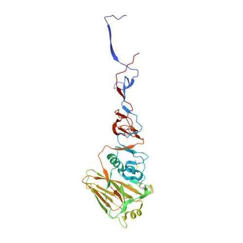
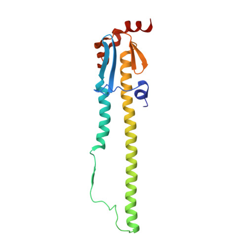
    In 2004 an hemagglutinin 3 neuraminidase 8 (H3N8) equine influenza virus was transmitted from horses to dogs in Florida and subsequently spread throughout the United States and to Europe. To understand the molecular basis of changes in the antigenicity of H3 hemagglutinins (HAs) that have occurred during virus evolution in horses, and to investigate the role of HA in the equine to canine cross-species transfer, we used X-ray crystallography to determine the structures of the HAs from two antigenically distinct equine viruses and from a canine virus. Structurally all three are very similar with the majority of amino acid sequence differences between the two equine HAs located on the virus membrane-distal molecular surface. HAs of canine viruses are distinct in containing a Trp-222 → Leu substitution in the receptor binding site that influences specificity for receptor analogs. In the fusion subdomain of canine and recent equine virus HAs a unique difference is observed by comparison with all other HAs examined to date. Analyses of site-specific mutant HAs indicate that a single amino acid substitution, Thr-30 → Ser, influences interactions between N-terminal and C-terminal regions of the subdomain that are important in the structural changes required for membrane fusion activity. Both structural modifications may have facilitated the transmission of H3N8 influenza from horses to dogs.


  • Organizational Affiliation

    Divisions of Virology,Molecular Structure, and.


Macromolecules
Find similar proteins by:  (by identity cutoff)  |  3D Structure
Entity ID: 1
MoleculeChains Sequence LengthOrganismDetailsImage
H3 HAEMAGGLUTININ HA1 CHAIN
A, C, E
327Influenza A virus (A/equine/Richmond/1/2007(H3N8))Mutation(s): 1 
UniProt
Find proteins for C3TUR9 (Influenza A virus)
Explore C3TUR9 
Go to UniProtKB:  C3TUR9
Entity Groups  
Sequence Clusters30% Identity50% Identity70% Identity90% Identity95% Identity100% Identity
UniProt GroupC3TUR9
Sequence Annotations
Expand
  • Reference Sequence
Find similar proteins by:  (by identity cutoff)  |  3D Structure
Entity ID: 2
MoleculeChains Sequence LengthOrganismDetailsImage
H3 HAEMAGGLUTININ HA2 CHAIN
B, D, F
172Influenza A virus (A/equine/Richmond/1/2007(H3N8))Mutation(s): 0 
UniProt
Find proteins for C3TUR9 (Influenza A virus)
Explore C3TUR9 
Go to UniProtKB:  C3TUR9
Entity Groups  
Sequence Clusters30% Identity50% Identity70% Identity90% Identity95% Identity100% Identity
UniProt GroupC3TUR9
Sequence Annotations
Expand
  • Reference Sequence
Oligosaccharides

Help

Entity ID: 3
MoleculeChains Length2D Diagram Glycosylation3D Interactions
beta-D-mannopyranose-(1-4)-2-acetamido-2-deoxy-beta-D-glucopyranose-(1-4)-[alpha-L-fucopyranose-(1-6)]2-acetamido-2-deoxy-beta-D-glucopyranose
G
4N-Glycosylation
Glycosylation Resources
GlyTouCan:  G32152BH
GlyCosmos:  G32152BH
GlyGen:  G32152BH
Entity ID: 4
MoleculeChains Length2D Diagram Glycosylation3D Interactions
alpha-D-mannopyranose-(1-3)-[alpha-D-mannopyranose-(1-6)]beta-D-mannopyranose-(1-4)-2-acetamido-2-deoxy-beta-D-glucopyranose-(1-4)-2-acetamido-2-deoxy-beta-D-glucopyranose
H, J, K
5N-Glycosylation
Glycosylation Resources
GlyTouCan:  G22768VO
GlyCosmos:  G22768VO
GlyGen:  G22768VO
Entity ID: 5
MoleculeChains Length2D Diagram Glycosylation3D Interactions
alpha-D-mannopyranose-(1-3)-[alpha-D-mannopyranose-(1-6)]beta-D-mannopyranose-(1-4)-2-acetamido-2-deoxy-beta-D-glucopyranose-(1-4)-[alpha-L-fucopyranose-(1-6)]2-acetamido-2-deoxy-beta-D-glucopyranose
I
6N-Glycosylation
Glycosylation Resources
GlyTouCan:  G82348BZ
GlyCosmos:  G82348BZ
GlyGen:  G82348BZ
Entity ID: 6
MoleculeChains Length2D Diagram Glycosylation3D Interactions
alpha-L-fucopyranose-(1-6)-2-acetamido-2-deoxy-beta-D-glucopyranose
L, O
2N-Glycosylation
Glycosylation Resources
GlyTouCan:  G86851RC
GlyCosmos:  G86851RC
GlyGen:  G86851RC
Entity ID: 7
MoleculeChains Length2D Diagram Glycosylation3D Interactions
2-acetamido-2-deoxy-beta-D-glucopyranose-(1-4)-2-acetamido-2-deoxy-beta-D-glucopyranose
M
2N-Glycosylation
Glycosylation Resources
GlyTouCan:  G42666HT
GlyCosmos:  G42666HT
GlyGen:  G42666HT
Entity ID: 8
MoleculeChains Length2D Diagram Glycosylation3D Interactions
beta-D-mannopyranose-(1-4)-2-acetamido-2-deoxy-beta-D-glucopyranose-(1-4)-2-acetamido-2-deoxy-beta-D-glucopyranose
N
3N-Glycosylation
Glycosylation Resources
GlyTouCan:  G15407YE
GlyCosmos:  G15407YE
GlyGen:  G15407YE
Small Molecules
Ligands 2 Unique
IDChains Name / Formula / InChI Key2D Diagram3D Interactions
NAG
Query on NAG

Download Ideal Coordinates CCD File 
P [auth A]
Q [auth A]
R [auth A]
T [auth B]
U [auth C]
P [auth A],
Q [auth A],
R [auth A],
T [auth B],
U [auth C],
V [auth E]
2-acetamido-2-deoxy-beta-D-glucopyranose
C8 H15 N O6
OVRNDRQMDRJTHS-FMDGEEDCSA-N
EDO
Query on EDO

Download Ideal Coordinates CCD File 
S [auth A],
W [auth E],
X [auth F]
1,2-ETHANEDIOL
C2 H6 O2
LYCAIKOWRPUZTN-UHFFFAOYSA-N
Experimental Data & Validation

Experimental Data

  • Method: X-RAY DIFFRACTION
  • Resolution: 2.87 Å
  • R-Value Free: 0.277 
  • R-Value Work: 0.217 
  • R-Value Observed: 0.220 
  • Space Group: P 21 21 21
Unit Cell:
Length ( Å )Angle ( ˚ )
a = 79.82α = 90
b = 129.62β = 90
c = 195.24γ = 90
Software Package:
Software NamePurpose
REFMACrefinement
XDSdata reduction
Aimlessdata scaling
PHASERphasing

Structure Validation

View Full Validation Report


Ligand Structure Quality Assessment 


Entry History 

Deposition Data

Revision History  (Full details and data files)

  • Version 1.0: 2014-07-23
    Type: Initial release
  • Version 1.1: 2014-08-13
    Changes: Database references
  • Version 2.0: 2020-07-29
    Type: Remediation
    Reason: Carbohydrate remediation
    Changes: Advisory, Atomic model, Data collection, Derived calculations, Other, Structure summary
  • Version 2.1: 2024-01-10
    Changes: Data collection, Database references, Refinement description, Structure summary