4UMM

The Cryo-EM structure of the palindromic DNA-bound USP-EcR nuclear receptor reveals an asymmetric organization with allosteric domain positioning


Experimental Data Snapshot

  • Method: ELECTRON MICROSCOPY
  • Resolution: 11.6 Å
  • Aggregation State: PARTICLE 
  • Reconstruction Method: SINGLE PARTICLE 

wwPDB Validation   3D Report Full Report


This is version 1.2 of the entry. See complete history


Literature

The Palindromic DNA-Bound Usp-Ecr Nuclear Receptor Adopts an Asymmetric Organization with Allosteric Domain Positioning.

Maletta, M.Orlov, I.Moras, D.Billas, I.M.L.Klaholz, B.P.

(2014) Nat Commun 5: 4139

  • DOI: https://doi.org/10.1038/ncomms5139
  • Primary Citation of Related Structures:  
    4UMM

  • PubMed Abstract: 

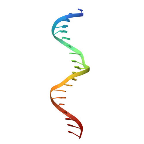
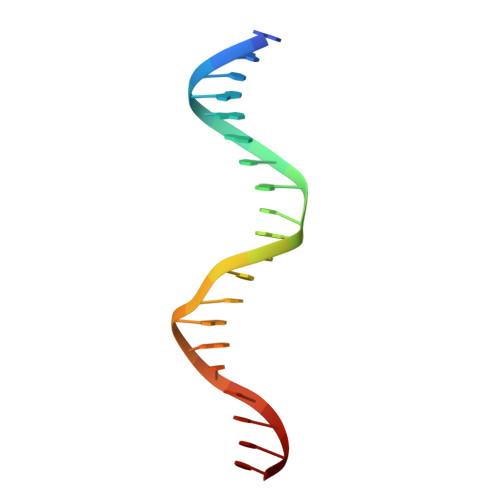
    Nuclear receptors (NRs) regulate gene expression through DNA- and ligand-binding and thus represent crucial therapeutic targets. The ultraspiracle protein/ecdysone receptor (USP/EcR) complex binds to half-sites with a one base pair spaced inverted repeat (IR1), a palindromic DNA response element (RE) reminiscent of IRs observed for vertebrate steroid hormone receptors. Here we present the cryo electron microscopy structure of the USP/EcR complex bound to an IR1 RE which provides the first description of a full IR-bound NR complex. The structure reveals that even though the DNA is almost symmetric, the complex adopts a highly asymmetric architecture in which the ligand-binding domains (LBDs) are positioned 5' off-centred. Additional interactions of the USP LBD with the 5'-flanking sequence trigger transcription activity as monitored by transfection assays. The comparison with DR-bound NR complexes suggests that DNA is the major allosteric driver in inversely positioning the LBDs, which serve as the main binding-site for transcriptional regulators.


  • Organizational Affiliation

    1] Centre for Integrative Biology (CBI), Department of Integrated Structural Biology, IGBMC (Institute of Genetics and of Molecular and Cellular Biology), 1 rue Laurent Fries, 67404 Illkirch, France [2] Centre National de la Recherche Scientifique (CNRS) UMR 7104, 67404 Illkirch, France [3] Institut National de la Santé et de la Recherche Médicale (INSERM) U964, 67404 Illkirch, France [4] Université de Strasbourg, 67404 Strasbourg, France.


Macromolecules

Find similar proteins by:  (by identity cutoff)  |  3D Structure
Entity ID: 1
MoleculeChains Sequence LengthOrganismDetailsImage
ECR-USP78Heliothis virescensMutation(s): 0 
Entity Groups  
Sequence Clusters30% Identity50% Identity70% Identity90% Identity95% Identity100% Identity
Sequence Annotations
Expand
  • Reference Sequence
Find similar proteins by:  (by identity cutoff)  |  3D Structure
Entity ID: 4
MoleculeChains Sequence LengthOrganismDetailsImage
ECDYSONE RECEPTORD [auth E]87Heliothis virescensMutation(s): 0 
UniProt
Find proteins for O18473 (Heliothis virescens)
Explore O18473 
Go to UniProtKB:  O18473
Entity Groups  
Sequence Clusters30% Identity50% Identity70% Identity90% Identity95% Identity100% Identity
UniProt GroupO18473
Sequence Annotations
Expand
  • Reference Sequence
Find similar proteins by:  (by identity cutoff)  |  3D Structure
Entity ID: 5
MoleculeChains Sequence LengthOrganismDetailsImage
GENE REGULATION PROTEINE [auth F]264Heliothis virescensMutation(s): 0 
UniProt
Find proteins for Q7SIF6 (Heliothis virescens)
Explore Q7SIF6 
Go to UniProtKB:  Q7SIF6
Entity Groups  
Sequence Clusters30% Identity50% Identity70% Identity90% Identity95% Identity100% Identity
UniProt GroupQ7SIF6
Sequence Annotations
Expand
  • Reference Sequence
Find similar proteins by:  (by identity cutoff)  |  3D Structure
Entity ID: 6
MoleculeChains Sequence LengthOrganismDetailsImage
ECDYSONE RECEPTORF [auth G]266Heliothis virescensMutation(s): 0 
UniProt
Find proteins for O18473 (Heliothis virescens)
Explore O18473 
Go to UniProtKB:  O18473
Entity Groups  
Sequence Clusters30% Identity50% Identity70% Identity90% Identity95% Identity100% Identity
UniProt GroupO18473
Sequence Annotations
Expand
  • Reference Sequence

Find similar nucleic acids by:  Sequence   |   3D Structure  

Entity ID: 2
MoleculeChains LengthOrganismImage
5'-D(*CP*AP*AP*GP*GP*GP*TP*TP*CP*AP*AP*TP*GP*CP *AP*CP*TP*TP*GP*TP)-3'B [auth C]20synthetic construct
Sequence Annotations
Expand
  • Reference Sequence

Find similar nucleic acids by:  Sequence   |   3D Structure  

Entity ID: 3
MoleculeChains LengthOrganismImage
5'-D(*DGP*AP*CP*AP*AP*GP*TP*GP*CP*AP*TP*TP*GP*DAP *AP*CP*CP*CP*TP*T)-3'C [auth D]20synthetic construct
Sequence Annotations
Expand
  • Reference Sequence
Small Molecules
Ligands 2 Unique
IDChains Name / Formula / InChI Key2D Diagram3D Interactions
EPH
Query on EPH

Download Ideal Coordinates CCD File 
G [auth F]L-ALPHA-PHOSPHATIDYL-BETA-OLEOYL-GAMMA-PALMITOYL-PHOSPHATIDYLETHANOLAMINE
C39 H68 N O8 P
MABRTXOVHMDVAT-AAEGOEIASA-N
P1A
Query on P1A

Download Ideal Coordinates CCD File 
H [auth G]2,3,14,20,22-PENTAHYDROXYCHOLEST-7-EN-6-ONE
C27 H44 O6
PJYYBCXMCWDUAZ-JJJZTNILSA-N
Binding Affinity Annotations 
IDSourceBinding Affinity
P1A BindingDB:  4UMM IC50: min: 1.29, max: 900 (nM) from 2 assay(s)
Experimental Data & Validation

Experimental Data

  • Method: ELECTRON MICROSCOPY
  • Resolution: 11.6 Å
  • Aggregation State: PARTICLE 
  • Reconstruction Method: SINGLE PARTICLE 
EM Software:
TaskSoftware PackageVersion
RECONSTRUCTIONIMAGIC5

Structure Validation

View Full Validation Report


Entry History 

Deposition Data

Revision History  (Full details and data files)

  • Version 1.0: 2014-06-25
    Type: Initial release
  • Version 1.1: 2014-07-02
    Changes: Database references, Other
  • Version 1.2: 2018-10-03
    Changes: Data collection, Derived calculations