Crystal structure of a DNA/Ba2+ G-quadruplex containing a water-mediated C-tetrad.
Zhang, D., Huang, T., Lukeman, P.S., Paukstelis, P.J.(2014) Nucleic Acids Res 42: 13422-13429
- PubMed: 25389267
- DOI: https://doi.org/10.1093/nar/gku1122
- Primary Citation of Related Structures:
4U92 - PubMed Abstract:
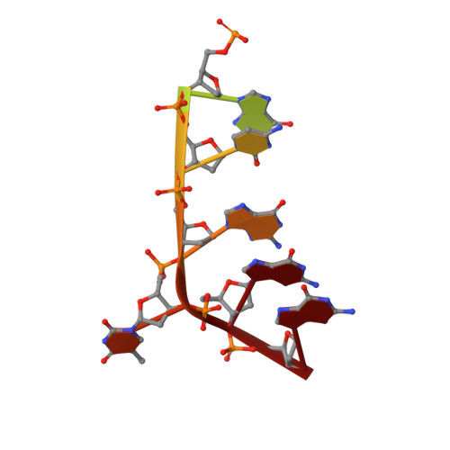
We have determined the 1.50 Å crystal structure of the DNA decamer, d(CCA(CNV)KGCGTGG) ((CNV)K, 3-cyanovinylcarbazole), which forms a G-quadruplex structure in the presence of Ba(2+). The structure contains several unique features including a bulged nucleotide and the first crystal structure observation of a C-tetrad. The structure reveals that water molecules mediate contacts between the divalent cations and the C-tetrad, allowing Ba(2+) ions to occupy adjacent steps in the central ion channel. One ordered Mg(2+) facilitates 3'-3' stacking of two quadruplexes in the asymmetric unit, while the bulged nucleotide mediates crystal contacts. Despite the high diffraction limit, the first four nucleotides including the (CNV)K nucleoside are disordered though they are still involved in crystal packing. This work suggests that the bulky hydrophobic groups may locally influence the formation of non-Watson-Crick structures from otherwise complementary sequences. These observations lead to the intriguing possibility that certain types of DNA damage may act as modulators of G-quadruplex formation.
Organizational Affiliation:
Department of Chemistry & Biochemistry, Center for Biomolecular Structure and Organization, Maryland NanoCenter, University of Maryland, College Park, MD 20742, USA.