


X-ray structure uridine phosphorylase from Vibrio cholerae in complex with new anticancer compound at 1.17 A resolution
Prokofev, I.I., Lashkov, A.A., Gabdoulkhakov, A.G., Betzel, C., Mikhailov, A.M.To be published.
Experimental Data Snapshot
Entity ID: 1 | |||||
|---|---|---|---|---|---|
| Molecule | Chains | Sequence Length | Organism | Details | Image |
| Uridine phosphorylase | 253 | Vibrio cholerae | Mutation(s): 0 Gene Names: udp, DN30_1909, VC39_02535, VC78_02550, VS27_10630, WG08_05660 EC: 2.4.2.3 | 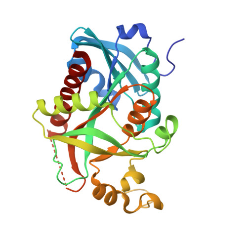 | |
UniProt | |||||
Find proteins for Q9K4U1 (Vibrio cholerae) Explore Q9K4U1 Go to UniProtKB: Q9K4U1 | |||||
Entity Groups | |||||
| Sequence Clusters | 30% Identity50% Identity70% Identity90% Identity95% Identity100% Identity | ||||
| UniProt Group | Q9K4U1 | ||||
Sequence AnnotationsExpand | |||||
| |||||
| Ligands 6 Unique | |||||
|---|---|---|---|---|---|
| ID | Chains | Name / Formula / InChI Key | 2D Diagram | 3D Interactions | |
| M5F Query on M5F | O [auth D], U [auth F] | 1-[(2S)-2,3-diaminopropyl]-5-fluoropyrimidine-2,4(1H,3H)-dione C7 H11 F N4 O2 ZBKWVMMCABYTAU-BYPYZUCNSA-N |  | ||
| 4WR Query on 4WR | J [auth B], P [auth D] | 1-[(2R)-2,3-diaminopropyl]-5-fluoropyrimidine-2,4(1H,3H)-dione C7 H11 F N4 O2 ZBKWVMMCABYTAU-SCSAIBSYSA-N |  | ||
| TRS Query on TRS | T [auth F] | 2-AMINO-2-HYDROXYMETHYL-PROPANE-1,3-DIOL C4 H12 N O3 LENZDBCJOHFCAS-UHFFFAOYSA-O |  | ||
| PEG Query on PEG | N [auth D] | DI(HYDROXYETHYL)ETHER C4 H10 O3 MTHSVFCYNBDYFN-UHFFFAOYSA-N |  | ||
| GOL Query on GOL | M [auth D], Q [auth E] | GLYCEROL C3 H8 O3 PEDCQBHIVMGVHV-UHFFFAOYSA-N |  | ||
| EDO Query on EDO | G [auth A] H [auth A] I [auth B] K [auth C] L [auth C] | 1,2-ETHANEDIOL C2 H6 O2 LYCAIKOWRPUZTN-UHFFFAOYSA-N |  | ||
| Length ( Å ) | Angle ( ˚ ) |
|---|---|
| a = 91.28 | α = 90 |
| b = 91.28 | β = 90 |
| c = 137.227 | γ = 120 |
| Software Name | Purpose |
|---|---|
| PHENIX | refinement |
| SCALA | data scaling |
| MOLREP | phasing |
| PDB_EXTRACT | data extraction |
| XDS | data reduction |
| XDS | data scaling |
RCSB PDB (citation) is hosted by
RCSB PDB is a member of the

