4TQU

Crystal structure of a bacterial ABC transporter involved in the import of the acidic polysaccharide alginate


Experimental Data Snapshot

  • Method: X-RAY DIFFRACTION
  • Resolution: 3.20 Å
  • R-Value Free: 0.282 
  • R-Value Work: 0.237 
  • R-Value Observed: 0.239 

wwPDB Validation   3D Report Full Report


This is version 2.0 of the entry. See complete history


Literature

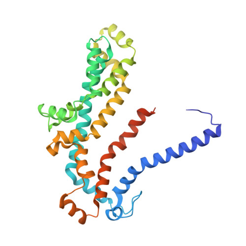
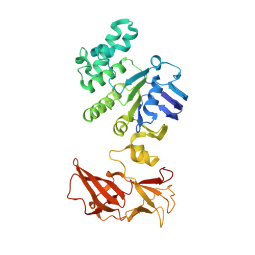
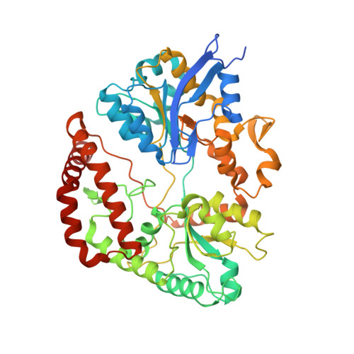
Structure of a Bacterial ABC Transporter Involved in the Import of an Acidic Polysaccharide Alginate

Maruyama, Y.Itoh, T.Kaneko, A.Nishitani, Y.Mikami, B.Hashimoto, W.Murata, K.

(2015) Structure 23: 1643-1654

  • DOI: https://doi.org/10.1016/j.str.2015.06.021
  • Primary Citation of Related Structures:  
    4TQU, 4TQV

  • PubMed Abstract: 

    The acidic polysaccharide alginate represents a promising marine biomass for the microbial production of biofuels, although the molecular and structural characteristics of alginate transporters remain to be clarified. In Sphingomonas sp. A1, the ATP-binding cassette transporter AlgM1M2SS is responsible for the import of alginate across the cytoplasmic membrane. Here, we present the substrate-transport characteristics and quaternary structure of AlgM1M2SS. The addition of poly- or oligoalginate enhanced the ATPase activity of reconstituted AlgM1M2SS coupled with one of the periplasmic solute-binding proteins, AlgQ1 or AlgQ2. External fluorescence-labeled oligoalginates were specifically imported into AlgM1M2SS-containing proteoliposomes in the presence of AlgQ2, ATP, and Mg(2+). The crystal structure of AlgQ2-bound AlgM1M2SS adopts an inward-facing conformation. The interaction between AlgQ2 and AlgM1M2SS induces the formation of an alginate-binding tunnel-like structure accessible to the solvent. The translocation route inside the transmembrane domains contains charged residues suitable for the import of acidic saccharides.


  • Organizational Affiliation

    Division of Food Science and Biotechnology, Graduate School of Agriculture, Kyoto University, Gokasho, Uji, Kyoto 611-0011, Japan.


Macromolecules
Find similar proteins by:  (by identity cutoff)  |  3D Structure
Entity ID: 1
MoleculeChains Sequence LengthOrganismDetailsImage
AlgM1A [auth M]301Sphingomonas sp.Mutation(s): 0 
Gene Names: algM1
Membrane Entity: Yes 
UniProt
Find proteins for Q9KWT8 (Sphingomonas sp)
Explore Q9KWT8 
Go to UniProtKB:  Q9KWT8
Entity Groups  
Sequence Clusters30% Identity50% Identity70% Identity90% Identity95% Identity100% Identity
UniProt GroupQ9KWT8
Sequence Annotations
Expand
  • Reference Sequence
Find similar proteins by:  (by identity cutoff)  |  3D Structure
Entity ID: 2
MoleculeChains Sequence LengthOrganismDetailsImage
AlgM2B [auth N]305Sphingomonas sp.Mutation(s): 0 
Gene Names: algM2
Membrane Entity: Yes 
UniProt
Find proteins for Q9KWT7 (Sphingomonas sp)
Explore Q9KWT7 
Go to UniProtKB:  Q9KWT7
Entity Groups  
Sequence Clusters30% Identity50% Identity70% Identity90% Identity95% Identity100% Identity
UniProt GroupQ9KWT7
Sequence Annotations
Expand
  • Reference Sequence
Find similar proteins by:  (by identity cutoff)  |  3D Structure
Entity ID: 3
MoleculeChains Sequence LengthOrganismDetailsImage
AlgSC [auth S],
E [auth T]
363Sphingomonas sp.Mutation(s): 1 
Gene Names: algS
UniProt
Find proteins for Q9KWT9 (Sphingomonas sp)
Explore Q9KWT9 
Go to UniProtKB:  Q9KWT9
Entity Groups  
Sequence Clusters30% Identity50% Identity70% Identity90% Identity95% Identity100% Identity
UniProt GroupQ9KWT9
Sequence Annotations
Expand
  • Reference Sequence
Find similar proteins by:  (by identity cutoff)  |  3D Structure
Entity ID: 4
MoleculeChains Sequence LengthOrganismDetailsImage
AlgQ2D [auth Q]516Sphingomonas sp.Mutation(s): 0 
Gene Names: algQ2
Membrane Entity: Yes 
UniProt
Find proteins for Q9KWT5 (Sphingomonas sp)
Explore Q9KWT5 
Go to UniProtKB:  Q9KWT5
Entity Groups  
Sequence Clusters30% Identity50% Identity70% Identity90% Identity95% Identity100% Identity
UniProt GroupQ9KWT5
Sequence Annotations
Expand
  • Reference Sequence
Oligosaccharides

Help

Entity ID: 5
MoleculeChains Length2D Diagram Glycosylation3D Interactions
4-deoxy-alpha-L-erythro-hex-4-enopyranuronic acid-(1-4)-beta-D-mannopyranuronic acid-(1-4)-beta-D-mannopyranuronic acid-(1-4)-beta-D-mannopyranuronic acidF [auth A]4N/A
Glycosylation Resources
GlyTouCan:  G76472TU
GlyCosmos:  G76472TU
Small Molecules
Ligands 1 Unique
IDChains Name / Formula / InChI Key2D Diagram3D Interactions
CA
Query on CA

Download Ideal Coordinates CCD File 
G [auth Q]CALCIUM ION
Ca
BHPQYMZQTOCNFJ-UHFFFAOYSA-N
Experimental Data & Validation

Experimental Data

  • Method: X-RAY DIFFRACTION
  • Resolution: 3.20 Å
  • R-Value Free: 0.282 
  • R-Value Work: 0.237 
  • R-Value Observed: 0.239 
  • Space Group: P 21 21 21
Unit Cell:
Length ( Å )Angle ( ˚ )
a = 71.367α = 90
b = 134.177β = 90
c = 273.798γ = 90
Software Package:
Software NamePurpose
PHENIXrefinement

Structure Validation

View Full Validation Report


Entry History & Funding Information

Deposition Data


Funding OrganizationLocationGrant Number
Grants-in-Aid for Scientific Research from Japan Society for the Promotion of ScienceJapan--
Targeted Proteins Research Program from Ministry of Education, Culture, Sports, Science, and TechnologyJapan--

Revision History  (Full details and data files)

  • Version 1.0: 2015-07-22
    Type: Initial release
  • Version 1.1: 2015-08-19
    Changes: Database references
  • Version 1.2: 2015-09-16
    Changes: Database references
  • Version 1.3: 2020-01-29
    Changes: Data collection, Database references, Derived calculations
  • Version 2.0: 2020-07-29
    Type: Remediation
    Reason: Carbohydrate remediation
    Changes: Advisory, Atomic model, Data collection, Derived calculations, Structure summary