4RSZ

The X-ray structure of the Primary Adduct formed in the Reaction between Cisplatin and Cytochrome c


Experimental Data Snapshot

  • Method: X-RAY DIFFRACTION
  • Resolution: 2.19 Å
  • R-Value Free: 0.282 
  • R-Value Work: 0.231 
  • R-Value Observed: 0.234 

wwPDB Validation   3D Report Full Report


Ligand Structure Quality Assessment 


This is version 2.1 of the entry. See complete history


Literature

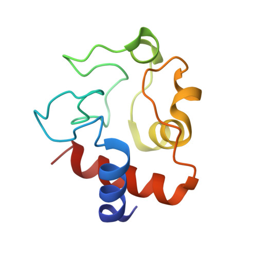
The X-ray structure of the primary adducts formed in the reaction between cisplatin and cytochrome c.

Ferraro, G.Messori, L.Merlino, A.

(2015) Chem Commun (Camb) 51: 2559-2561

  • DOI: https://doi.org/10.1039/c4cc09056j
  • Primary Citation of Related Structures:  
    4RSZ

  • PubMed Abstract: 

    In the present study, the interactions between cisplatin and cytochrome c are investigated. Based on high-resolution X-ray diffraction data, two monometalated species, i.e. cyt c-Pt(NH3)2 and cyt c-Pt(NH3)2Cl, are found to be the main adducts that form in the reaction between the protein and the drug. Both monodentate and bidentate platinum coordination to the protein is observed, with platinum binding either to Met65 or to Met65 and Glu61, simultaneously.


  • Organizational Affiliation

    Department of Chemical Sciences, University of Naples Federico II, Complesso Universitario di Monte Sant'Angelo, Via Cintia, I-80126, Napoli, Italy. antonello.merlino@unina.it.


Macromolecules
Find similar proteins by:  (by identity cutoff)  |  3D Structure
Entity ID: 1
MoleculeChains Sequence LengthOrganismDetailsImage
Cytochrome c
A, B, C, D, E
A, B, C, D, E, F
104Equus caballusMutation(s): 0 
UniProt
Find proteins for P00004 (Equus caballus)
Explore P00004 
Go to UniProtKB:  P00004
Entity Groups  
Sequence Clusters30% Identity50% Identity70% Identity90% Identity95% Identity100% Identity
UniProt GroupP00004
Sequence Annotations
Expand
  • Reference Sequence
Small Molecules
Ligands 4 Unique
IDChains Name / Formula / InChI Key2D Diagram3D Interactions
HEC
Query on HEC

Download Ideal Coordinates CCD File 
EA [auth F]
G [auth A]
K [auth B]
Q [auth C]
V [auth D]
EA [auth F],
G [auth A],
K [auth B],
Q [auth C],
V [auth D],
Z [auth E]
HEME C
C34 H34 Fe N4 O4
HXQIYSLZKNYNMH-LJNAALQVSA-N
CPT
Query on CPT

Download Ideal Coordinates CCD File 
AA [auth E],
GA [auth F],
H [auth A],
M [auth B],
W [auth D]
Cisplatin
Cl2 H6 N2 Pt
LXZZYRPGZAFOLE-UHFFFAOYSA-L
SO4
Query on SO4

Download Ideal Coordinates CCD File 
FA [auth F],
L [auth B]
SULFATE ION
O4 S
QAOWNCQODCNURD-UHFFFAOYSA-L
NO3
Query on NO3

Download Ideal Coordinates CCD File 
BA [auth E]
CA [auth E]
DA [auth E]
HA [auth F]
I [auth A]
BA [auth E],
CA [auth E],
DA [auth E],
HA [auth F],
I [auth A],
IA [auth F],
J [auth A],
N [auth B],
O [auth B],
P [auth B],
R [auth C],
S [auth C],
T [auth C],
U [auth C],
X [auth D],
Y [auth D]
NITRATE ION
N O3
NHNBFGGVMKEFGY-UHFFFAOYSA-N
Experimental Data & Validation

Experimental Data

  • Method: X-RAY DIFFRACTION
  • Resolution: 2.19 Å
  • R-Value Free: 0.282 
  • R-Value Work: 0.231 
  • R-Value Observed: 0.234 
  • Space Group: P 3
Unit Cell:
Length ( Å )Angle ( ˚ )
a = 120.344α = 90
b = 120.344β = 90
c = 36.673γ = 120
Software Package:
Software NamePurpose
CrystalCleardata collection
PHASERphasing
REFMACrefinement
HKL-2000data reduction
HKL-2000data scaling

Structure Validation

View Full Validation Report


Ligand Structure Quality Assessment 


Entry History 

Deposition Data

  • Released Date: 2015-01-14 
  • Deposition Author(s): Merlino, A.

Revision History  (Full details and data files)

  • Version 1.0: 2015-01-14
    Type: Initial release
  • Version 1.1: 2015-02-25
    Changes: Database references
  • Version 2.0: 2021-03-10
    Changes: Advisory, Atomic model, Data collection, Derived calculations, Non-polymer description, Structure summary
  • Version 2.1: 2023-09-20
    Changes: Data collection, Database references, Refinement description