4RGA

Phage 1358 receptor binding protein in complex with the trisaccharide GlcNAc-Galf-GlcOMe


Experimental Data Snapshot

  • Method: X-RAY DIFFRACTION
  • Resolution: 2.10 Å
  • R-Value Free: 0.192 
  • R-Value Work: 0.180 
  • R-Value Observed: 0.181 

wwPDB Validation   3D Report Full Report


This is version 2.1 of the entry. See complete history


Literature

The targeted recognition of Lactococcus lactis phages to their polysaccharide receptors.

McCabe, O.Spinelli, S.Farenc, C.Labbe, M.Tremblay, D.Blangy, S.Oscarson, S.Moineau, S.Cambillau, C.

(2015) Mol Microbiol 96: 875-886

  • DOI: https://doi.org/10.1111/mmi.12978
  • Primary Citation of Related Structures:  
    4RGA

  • PubMed Abstract: 

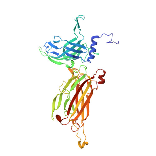
    Each phage infects a limited number of bacterial strains through highly specific interactions of the receptor-binding protein (RBP) at the tip of phage tail and the receptor at the bacterial surface. Lactococcus lactis is covered with a thin polysaccharide pellicle (hexasaccharide repeating units), which is used by a subgroup of phages as a receptor. Using L. lactis and phage 1358 as a model, we investigated the interaction between the phage RBP and the pellicle hexasaccharide of the host strain. A core trisaccharide (TriS), derived from the pellicle hexasaccharide repeating unit, was chemically synthesised, and the crystal structure of the RBP/TriS complex was determined. This provided unprecedented structural details of RBP/receptor site-specific binding. The complete hexasaccharide repeating unit was modelled and found to aptly fit the extended binding site. The specificity observed in in vivo phage adhesion assays could be interpreted in view of the reported structure. Therefore, by combining synthetic carbohydrate chemistry, X-ray crystallography and phage plaquing assays, we suggest that phage adsorption results from distinct recognition of the RBP towards the core TriS or the remaining residues of the hexasacchride receptor. This study provides a novel insight into the adsorption process of phages targeting saccharides as their receptors.


  • Organizational Affiliation

    Centre for Molecular Innovation and Drug Discovery, School of Chemistry and Chemical Biology, University College Dublin, Belfield, Dublin, Ireland.


Macromolecules
Find similar proteins by:  (by identity cutoff)  |  3D Structure
Entity ID: 1
MoleculeChains Sequence LengthOrganismDetailsImage
Phage 1358 receptor binding protein (ORF20)
A, B
393Lactococcus phage 1358Mutation(s): 0 
Gene Names: ORF20
UniProt
Find proteins for D3W0F1 (Lactococcus phage 1358)
Explore D3W0F1 
Go to UniProtKB:  D3W0F1
Entity Groups  
Sequence Clusters30% Identity50% Identity70% Identity90% Identity95% Identity100% Identity
UniProt GroupD3W0F1
Sequence Annotations
Expand
  • Reference Sequence
Oligosaccharides

Help

Entity ID: 2
MoleculeChains Length2D Diagram Glycosylation3D Interactions
2-acetamido-2-deoxy-beta-D-glucopyranose-(1-2)-beta-D-galactofuranose-(1-6)-methyl alpha-D-glucopyranoside
C, D
3N/A
Glycosylation Resources
GlyTouCan:  G42450WO
GlyCosmos:  G42450WO
Experimental Data & Validation

Experimental Data

  • Method: X-RAY DIFFRACTION
  • Resolution: 2.10 Å
  • R-Value Free: 0.192 
  • R-Value Work: 0.180 
  • R-Value Observed: 0.181 
  • Space Group: P 21 3
Unit Cell:
Length ( Å )Angle ( ˚ )
a = 166.37α = 90
b = 166.37β = 90
c = 166.37γ = 90
Software Package:
Software NamePurpose
PROTEUM PLUSdata collection
MOLREPphasing
BUSTERrefinement
XDSdata reduction
XSCALEdata scaling

Structure Validation

View Full Validation Report


Entry History 

Deposition Data

Revision History  (Full details and data files)

  • Version 1.0: 2015-05-20
    Type: Initial release
  • Version 2.0: 2020-07-29
    Type: Remediation
    Reason: Carbohydrate remediation
    Changes: Atomic model, Data collection, Database references, Derived calculations, Non-polymer description, Structure summary
  • Version 2.1: 2023-09-20
    Changes: Data collection, Database references, Refinement description, Structure summary