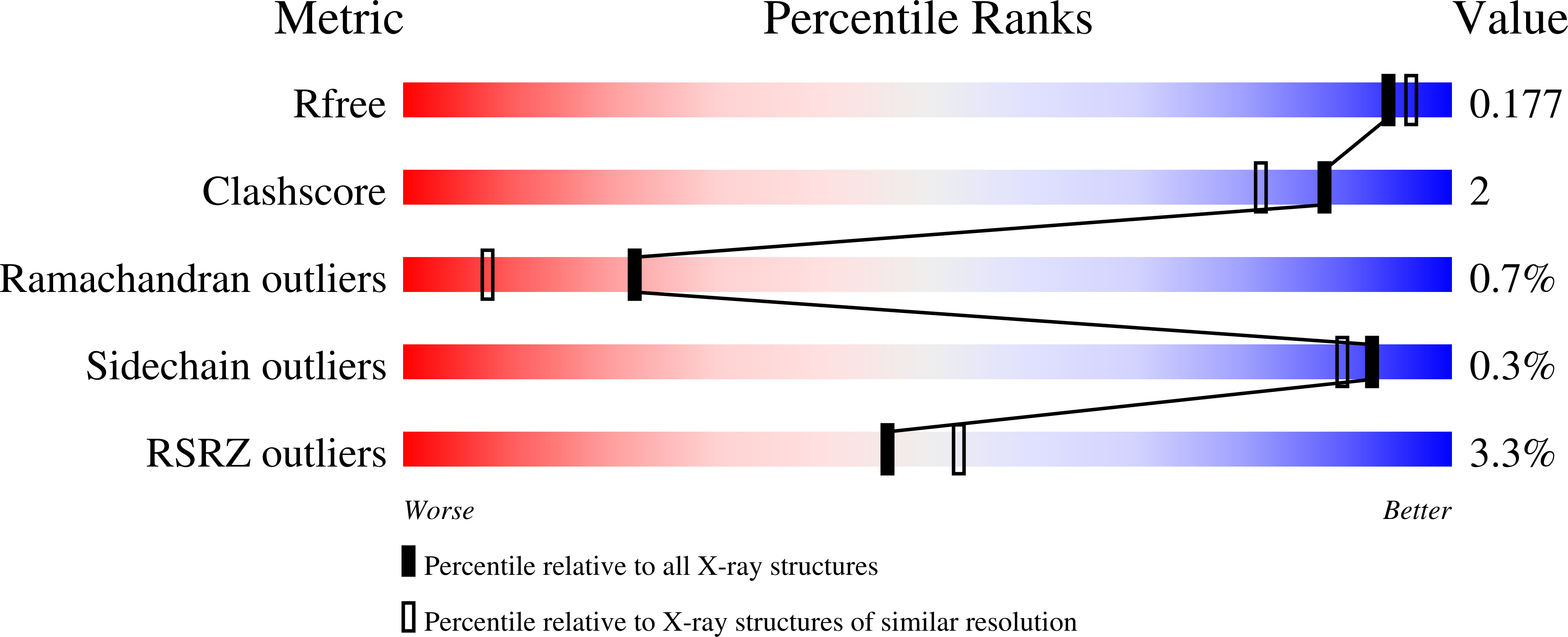
4RBM

Porphyromonas gingivalis gingipain K (Kgp) catalytic and immunoglobulin superfamily-like domains


Experimental Data Snapshot

  • Method: X-RAY DIFFRACTION
  • Resolution: 1.75 Å
  • R-Value Free: 0.172 
  • R-Value Work: 0.149 
  • R-Value Observed: 0.150 

wwPDB Validation   3D Report Full Report


Ligand Structure Quality Assessment 


This is version 2.0 of the entry. See complete history


Literature

Structure and Mechanism of Cysteine Peptidase Gingipain K (Kgp), a Major Virulence Factor of Porphyromonas gingivalis in Periodontitis.

de Diego, I.Veillard, F.Sztukowska, M.N.Guevara, T.Potempa, B.Pomowski, A.Huntington, J.A.Potempa, J.Gomis-Ruth, F.X.

(2014) J Biol Chem 289: 32291-32302

  • DOI: https://doi.org/10.1074/jbc.M114.602052
  • Primary Citation of Related Structures:  
    4RBM

  • PubMed Abstract: 

    Cysteine peptidases are key proteolytic virulence factors of the periodontopathogen Porphyromonas gingivalis, which causes chronic periodontitis, the most prevalent dysbiosis-driven disease in humans. Two peptidases, gingipain K (Kgp) and R (RgpA and RgpB), which differ in their selectivity after lysines and arginines, respectively, collectively account for 85% of the extracellular proteolytic activity of P. gingivalis at the site of infection. Therefore, they are promising targets for the design of specific inhibitors. Although the structure of the catalytic domain of RgpB is known, little is known about Kgp, which shares only 27% sequence identity. We report the high resolution crystal structure of a competent fragment of Kgp encompassing the catalytic cysteine peptidase domain and a downstream immunoglobulin superfamily-like domain, which is required for folding and secretion of Kgp in vivo. The structure, which strikingly resembles a tooth, was serendipitously trapped with a fragment of a covalent inhibitor targeting the catalytic cysteine. This provided accurate insight into the active site and suggested that catalysis may require a catalytic triad, Cys(477)-His(444)-Asp(388), rather than the cysteine-histidine dyad normally found in cysteine peptidases. In addition, a 20-Å-long solvent-filled interior channel traverses the molecule and links the bottom of the specificity pocket with the molecular surface opposite the active site cleft. This channel, absent in RgpB, may enhance the plasticity of the enzyme, which would explain the much lower activity in vitro toward comparable specific synthetic substrates. Overall, the present results report the architecture and molecular determinants of the working mechanism of Kgp, including interaction with its substrates.


  • Organizational Affiliation

    Proteolysis Lab, Molecular Biology Institute of Barcelona, Spanish Research Council (Consejo Superior de Investigaciones Cientificas), Barcelona Science Park, Helix Building, Baldiri Reixac 15-21, 08028 Barcelona, Catalonia, Spain.


Macromolecules
Find similar proteins by:  (by identity cutoff)  |  3D Structure
Entity ID: 1
MoleculeChains Sequence LengthOrganismDetailsImage
Lys-gingipain W83461Porphyromonas gingivalisMutation(s): 0 
Gene Names: kgpprtKprtP
EC: 3.4.22.47
UniProt
Find proteins for Q51817 (Porphyromonas gingivalis)
Explore Q51817 
Go to UniProtKB:  Q51817
Entity Groups  
Sequence Clusters30% Identity50% Identity70% Identity90% Identity95% Identity100% Identity
UniProt GroupQ51817
Sequence Annotations
Expand
  • Reference Sequence
Small Molecules
Ligands 8 Unique
IDChains Name / Formula / InChI Key2D Diagram3D Interactions
HIS
Query on HIS

Download Ideal Coordinates CCD File 
H [auth A]HISTIDINE
C6 H10 N3 O2
HNDVDQJCIGZPNO-YFKPBYRVSA-O
CKC
Query on CKC

Download Ideal Coordinates CCD File 
R [auth A](3S)-3,7-diaminoheptan-2-one
C7 H16 N2 O
FADKJNSSIWTVAI-ZETCQYMHSA-N
GOL
Query on GOL

Download Ideal Coordinates CCD File 
N [auth A],
O [auth A],
P [auth A],
Q [auth A]
GLYCEROL
C3 H8 O3
PEDCQBHIVMGVHV-UHFFFAOYSA-N
ACT
Query on ACT

Download Ideal Coordinates CCD File 
I [auth A],
J [auth A]
ACETATE ION
C2 H3 O2
QTBSBXVTEAMEQO-UHFFFAOYSA-M
NI
Query on NI

Download Ideal Coordinates CCD File 
F [auth A]NICKEL (II) ION
Ni
VEQPNABPJHWNSG-UHFFFAOYSA-N
AZI
Query on AZI

Download Ideal Coordinates CCD File 
K [auth A],
L [auth A],
M [auth A]
AZIDE ION
N3
IVRMZWNICZWHMI-UHFFFAOYSA-N
CA
Query on CA

Download Ideal Coordinates CCD File 
B [auth A],
C [auth A]
CALCIUM ION
Ca
BHPQYMZQTOCNFJ-UHFFFAOYSA-N
NA
Query on NA

Download Ideal Coordinates CCD File 
D [auth A],
E [auth A],
G [auth A]
SODIUM ION
Na
FKNQFGJONOIPTF-UHFFFAOYSA-N
Experimental Data & Validation

Experimental Data

  • Method: X-RAY DIFFRACTION
  • Resolution: 1.75 Å
  • R-Value Free: 0.172 
  • R-Value Work: 0.149 
  • R-Value Observed: 0.150 
  • Space Group: P 21 21 21
Unit Cell:
Length ( Å )Angle ( ˚ )
a = 56.64α = 90
b = 58.81β = 90
c = 135.5γ = 90
Software Package:
Software NamePurpose
ADSCdata collection
PHASERphasing
BUSTERrefinement
XDSdata reduction
XDSdata scaling
XSCALEdata scaling

Structure Validation

View Full Validation Report


Ligand Structure Quality Assessment 


Entry History 

Deposition Data

Revision History  (Full details and data files)

  • Version 1.0: 2014-10-08
    Type: Initial release
  • Version 1.1: 2014-10-15
    Changes: Database references
  • Version 1.2: 2014-12-10
    Changes: Database references
  • Version 1.3: 2017-11-22
    Changes: Refinement description
  • Version 1.4: 2023-09-20
    Changes: Data collection, Database references, Derived calculations, Refinement description
  • Version 2.0: 2023-11-15
    Changes: Atomic model, Data collection, Derived calculations