


Crystal structure of 5-methylcytosine deaminase from Klebsiella pneumoniae liganded with phosphonocytosine
Fedorov, A.A., Fedorov, E.V., Hitchcock, D.S., Raushel, F.M., Almo, S.C.To be published.
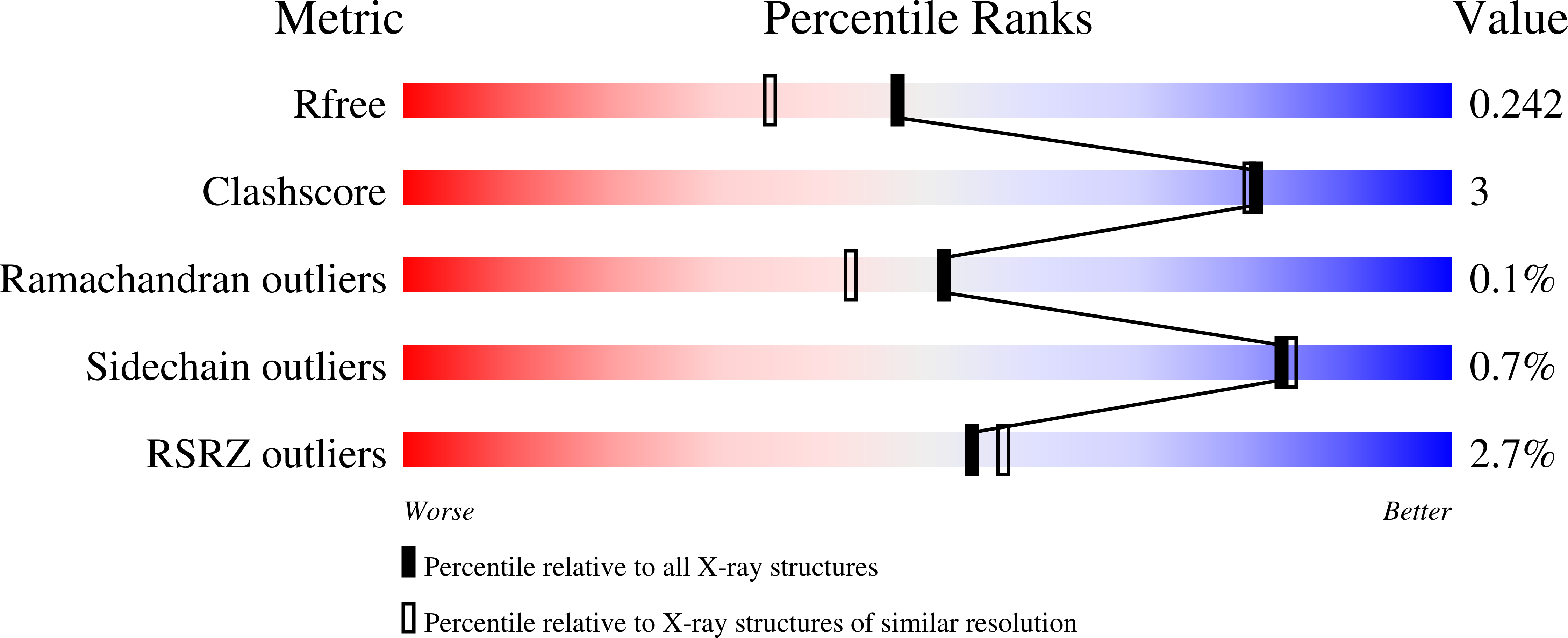
Experimental Data Snapshot
wwPDB Validation 3D Report Full Report
Entity ID: 1 | |||||
|---|---|---|---|---|---|
| Molecule | Chains | Sequence Length | Organism | Details | Image |
| Cytosine deaminase | 431 | Klebsiella pneumoniae 30660/NJST258_1 | Mutation(s): 0 Gene Names: KPNJ1_03949 EC: 3.5.4.1 | 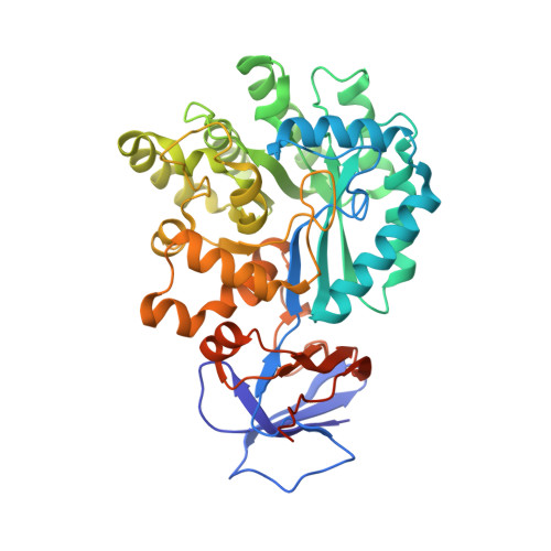 | |
UniProt | |||||
Find proteins for A0A0E1CHI1 (Klebsiella pneumoniae 30660/NJST258_1) Explore A0A0E1CHI1 Go to UniProtKB: A0A0E1CHI1 | |||||
Entity Groups | |||||
| Sequence Clusters | 30% Identity50% Identity70% Identity90% Identity95% Identity100% Identity | ||||
| UniProt Group | A0A0E1CHI1 | ||||
Sequence AnnotationsExpand | |||||
| |||||
| Ligands 5 Unique | |||||
|---|---|---|---|---|---|
| ID | Chains | Name / Formula / InChI Key | 2D Diagram | 3D Interactions | |
| O7U Query on O7U | DA [auth F] H [auth A] M [auth B] P [auth C] V [auth D] | (2R)-2-amino-2,5-dihydro-1,5,2-diazaphosphinin-6(1H)-one 2-oxide C3 H6 N3 O2 P GGLLBAYBJJLFCT-SECBINFHSA-N |  | ||
| PEG Query on PEG | BA [auth E], I [auth A], R [auth C], S [auth C], W [auth D] | DI(HYDROXYETHYL)ETHER C4 H10 O3 MTHSVFCYNBDYFN-UHFFFAOYSA-N |  | ||
| GOL Query on GOL | Q [auth C] | GLYCEROL C3 H8 O3 PEDCQBHIVMGVHV-UHFFFAOYSA-N |  | ||
| EDO Query on EDO | AA [auth E] J [auth A] K [auth A] N [auth B] T [auth C] | 1,2-ETHANEDIOL C2 H6 O2 LYCAIKOWRPUZTN-UHFFFAOYSA-N |  | ||
| FE2 Query on FE2 | CA [auth F] G [auth A] L [auth B] O [auth C] U [auth D] | FE (II) ION Fe CWYNVVGOOAEACU-UHFFFAOYSA-N |  | ||
| Length ( Å ) | Angle ( ˚ ) |
|---|---|
| a = 102.167 | α = 90 |
| b = 147.808 | β = 90 |
| c = 185.336 | γ = 90 |
| Software Name | Purpose |
|---|---|
| CBASS | data collection |
| BALBES | phasing |
| PHENIX | refinement |
| HKL-2000 | data reduction |
| HKL-2000 | data scaling |
RCSB PDB (citation) is hosted by
RCSB PDB is a member of the

