


Crystal Structure of Amidohydrolase Pmi1525 from Proteus Mirabilis Hi4320
Patskovsky, Y., Toro, R., Xiang, D.F., Raushel, F.M., Almo, S.C.To be published.
Experimental Data Snapshot
wwPDB Validation 3D Report Full Report

Entity ID: 1 | |||||
|---|---|---|---|---|---|
| Molecule | Chains | Sequence Length | Organism | Details | Image |
| Amidohydrolase Pmi1525 | 365 | Proteus mirabilis HI4320 | Mutation(s): 0 Gene Names: PMI1525 | 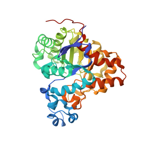 | |
UniProt | |||||
Find proteins for B4EXV8 (Proteus mirabilis (strain HI4320)) Explore B4EXV8 Go to UniProtKB: B4EXV8 | |||||
Entity Groups | |||||
| Sequence Clusters | 30% Identity50% Identity70% Identity90% Identity95% Identity100% Identity | ||||
| UniProt Group | B4EXV8 | ||||
Sequence AnnotationsExpand | |||||
| |||||
| Ligands 3 Unique | |||||
|---|---|---|---|---|---|
| ID | Chains | Name / Formula / InChI Key | 2D Diagram | 3D Interactions | |
| SO4 Query on SO4 | E [auth A], F [auth A] | SULFATE ION O4 S QAOWNCQODCNURD-UHFFFAOYSA-L |  | ||
| BUA Query on BUA | D [auth A] | butanoic acid C4 H8 O2 FERIUCNNQQJTOY-UHFFFAOYSA-N |  | ||
| MN Query on MN | B [auth A], C [auth A] | MANGANESE (II) ION Mn WAEMQWOKJMHJLA-UHFFFAOYSA-N |  | ||
| Length ( Å ) | Angle ( ˚ ) |
|---|---|
| a = 101.269 | α = 90 |
| b = 101.269 | β = 90 |
| c = 65.493 | γ = 120 |
| Software Name | Purpose |
|---|---|
| PHASER | phasing |
| REFMAC | refinement |
| HKL-3000 | data reduction |
| HKL-3000 | data scaling |
RCSB PDB (citation) is hosted by
RCSB PDB is a member of the

