


Crystal structures and reaction mechanisms of nitroalkane oxidase (NAO) from Pseudomonas aeruginosa
Chi, Y.M., Lee, J.H.To be published.
Experimental Data Snapshot
Entity ID: 1 | |||||
|---|---|---|---|---|---|
| Molecule | Chains | Sequence Length | Organism | Details | Image |
| Nitronate monooxygenase family protein | 356 | Pseudomonas aeruginosa PAO581 | Mutation(s): 0 Gene Names: M801_4200 EC: 1.7.3.1 | 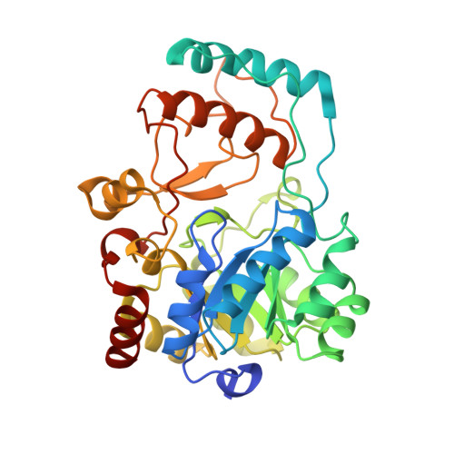 | |
UniProt | |||||
Find proteins for A0A0M3KKW0 (Pseudomonas aeruginosa PAO581) Explore A0A0M3KKW0 Go to UniProtKB: A0A0M3KKW0 | |||||
Entity Groups | |||||
| Sequence Clusters | 30% Identity50% Identity70% Identity90% Identity95% Identity100% Identity | ||||
| UniProt Group | A0A0M3KKW0 | ||||
Sequence AnnotationsExpand | |||||
| |||||
| Ligands 1 Unique | |||||
|---|---|---|---|---|---|
| ID | Chains | Name / Formula / InChI Key | 2D Diagram | 3D Interactions | |
| FMN Query on FMN | C [auth A], D [auth B] | FLAVIN MONONUCLEOTIDE C17 H21 N4 O9 P FVTCRASFADXXNN-SCRDCRAPSA-N |  | ||
| Length ( Å ) | Angle ( ˚ ) |
|---|---|
| a = 69.895 | α = 90 |
| b = 54.834 | β = 95.79 |
| c = 88.202 | γ = 90 |
| Software Name | Purpose |
|---|---|
| HKL-2000 | data collection |
| PHENIX | model building |
| PHENIX | refinement |
| HKL-2000 | data reduction |
| HKL-2000 | data scaling |
| PHENIX | phasing |
RCSB PDB (citation) is hosted by
RCSB PDB is a member of the

