4QFR

Structure of AMPK in complex with Cl-A769662 activator and STAUROSPORINE inhibitor


Experimental Data Snapshot

  • Method: X-RAY DIFFRACTION
  • Resolution: 3.34 Å
  • R-Value Free: 0.249 
  • R-Value Work: 0.210 
  • R-Value Observed: 0.212 

wwPDB Validation   3D Report Full Report


Ligand Structure Quality Assessment 


This is version 1.4 of the entry. See complete history


Literature

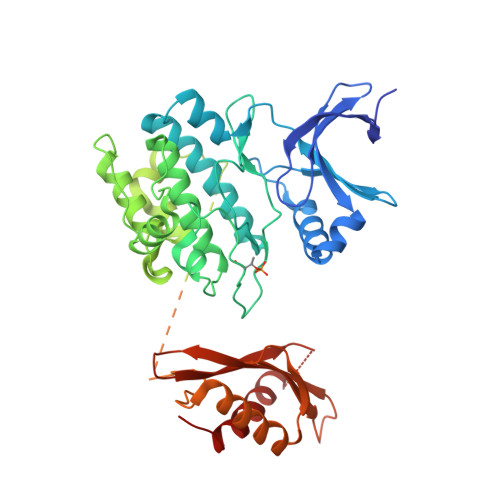
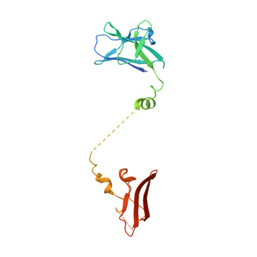
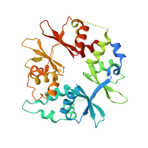
Structural Basis for AMPK Activation: Natural and Synthetic Ligands Regulate Kinase Activity from Opposite Poles by Different Molecular Mechanisms.

Calabrese, M.F.Rajamohan, F.Harris, M.S.Caspers, N.L.Magyar, R.Withka, J.M.Wang, H.Borzilleri, K.A.Sahasrabudhe, P.V.Hoth, L.R.Geoghegan, K.F.Han, S.Brown, J.Subashi, T.A.Reyes, A.R.Frisbie, R.K.Ward, J.Miller, R.A.Landro, J.A.Londregan, A.T.Carpino, P.A.Cabral, S.Smith, A.C.Conn, E.L.Cameron, K.O.Qiu, X.Kurumbail, R.G.

(2014) Structure 22: 1161-1172

  • DOI: https://doi.org/10.1016/j.str.2014.06.009
  • Primary Citation of Related Structures:  
    4QFG, 4QFR, 4QFS

  • PubMed Abstract: 

    AMP-activated protein kinase (AMPK) is a principal metabolic regulator affecting growth and response to cellular stress. Comprised of catalytic and regulatory subunits, each present in multiple forms, AMPK is best described as a family of related enzymes. In recent years, AMPK has emerged as a desirable target for modulation of numerous diseases, yet clinical therapies remain elusive. Challenges result, in part, from an incomplete understanding of the structure and function of full-length heterotrimeric complexes. In this work, we provide the full-length structure of the widely expressed α1β1γ1 isoform of mammalian AMPK, along with detailed kinetic and biophysical characterization. We characterize binding of the broadly studied synthetic activator A769662 and its analogs. Our studies follow on the heels of the recent disclosure of the α2β1γ1 structure and provide insight into the distinct molecular mechanisms of AMPK regulation by AMP and A769662.


  • Organizational Affiliation

    Worldwide Research and Development, Pfizer Inc., Eastern Point Road, Groton, CT 06340, USA.


Macromolecules
Find similar proteins by:  (by identity cutoff)  |  3D Structure
Entity ID: 1
MoleculeChains Sequence LengthOrganismDetailsImage
5'-AMP-activated protein kinase catalytic subunit alpha-1503Rattus norvegicusMutation(s): 0 
Gene Names: Ampk1Prkaa1
EC: 2.7.11.1 (PDB Primary Data), 2.7.11.27 (PDB Primary Data), 2.7.11.31 (PDB Primary Data), 2.7.11.26 (PDB Primary Data)
UniProt
Find proteins for P54645 (Rattus norvegicus)
Explore P54645 
Go to UniProtKB:  P54645
Entity Groups  
Sequence Clusters30% Identity50% Identity70% Identity90% Identity95% Identity100% Identity
UniProt GroupP54645
Sequence Annotations
Expand
  • Reference Sequence
Find similar proteins by:  (by identity cutoff)  |  3D Structure
Entity ID: 2
MoleculeChains Sequence LengthOrganismDetailsImage
5'-AMP-activated protein kinase subunit beta-1204Rattus norvegicusMutation(s): 1 
Gene Names: Prkab1
UniProt
Find proteins for P80386 (Rattus norvegicus)
Explore P80386 
Go to UniProtKB:  P80386
Entity Groups  
Sequence Clusters30% Identity50% Identity70% Identity90% Identity95% Identity100% Identity
UniProt GroupP80386
Sequence Annotations
Expand
  • Reference Sequence
Find similar proteins by:  (by identity cutoff)  |  3D Structure
Entity ID: 3
MoleculeChains Sequence LengthOrganismDetailsImage
5'-AMP-activated protein kinase subunit gamma-1330Rattus norvegicusMutation(s): 0 
Gene Names: Prkag1
UniProt
Find proteins for P80385 (Rattus norvegicus)
Explore P80385 
Go to UniProtKB:  P80385
Entity Groups  
Sequence Clusters30% Identity50% Identity70% Identity90% Identity95% Identity100% Identity
UniProt GroupP80385
Sequence Annotations
Expand
  • Reference Sequence
Small Molecules
Ligands 6 Unique
IDChains Name / Formula / InChI Key2D Diagram3D Interactions
STU
Query on STU

Download Ideal Coordinates CCD File 
D [auth A]STAUROSPORINE
C28 H26 N4 O3
HKSZLNNOFSGOKW-FYTWVXJKSA-N
ADP
Query on ADP

Download Ideal Coordinates CCD File 
M [auth C]ADENOSINE-5'-DIPHOSPHATE
C10 H15 N5 O10 P2
XTWYTFMLZFPYCI-KQYNXXCUSA-N
32J
Query on 32J

Download Ideal Coordinates CCD File 
I [auth A]2-chloro-4-hydroxy-3-(2'-hydroxybiphenyl-4-yl)-6-oxo-6,7-dihydrothieno[2,3-b]pyridine-5-carbonitrile
C20 H11 Cl N2 O3 S
JGMADXMPAZATOC-UHFFFAOYSA-N
AMP
Query on AMP

Download Ideal Coordinates CCD File 
K [auth C],
L [auth C]
ADENOSINE MONOPHOSPHATE
C10 H14 N5 O7 P
UDMBCSSLTHHNCD-KQYNXXCUSA-N
SO4
Query on SO4

Download Ideal Coordinates CCD File 
N [auth C]SULFATE ION
O4 S
QAOWNCQODCNURD-UHFFFAOYSA-L
CL
Query on CL

Download Ideal Coordinates CCD File 
E [auth A],
F [auth A],
G [auth A],
H [auth A],
J [auth B]
CHLORIDE ION
Cl
VEXZGXHMUGYJMC-UHFFFAOYSA-M
Modified Residues  1 Unique
IDChains TypeFormula2D DiagramParent
TPO
Query on TPO
A
L-PEPTIDE LINKINGC4 H10 N O6 PTHR
Binding Affinity Annotations 
IDSourceBinding Affinity
32J BindingDB:  4QFR Kd: 74 (nM) from 1 assay(s)
Experimental Data & Validation

Experimental Data

  • Method: X-RAY DIFFRACTION
  • Resolution: 3.34 Å
  • R-Value Free: 0.249 
  • R-Value Work: 0.210 
  • R-Value Observed: 0.212 
  • Space Group: P 61 2 2
Unit Cell:
Length ( Å )Angle ( ˚ )
a = 124.62α = 90
b = 124.62β = 90
c = 402.92γ = 120
Software Package:
Software NamePurpose
DENZOdata reduction
SCALEPACKdata scaling
PHASERphasing
BUSTER-TNTrefinement
PDB_EXTRACTdata extraction
BUSTERrefinement

Structure Validation

View Full Validation Report


Ligand Structure Quality Assessment 


Entry History 

Deposition Data

Revision History  (Full details and data files)

  • Version 1.0: 2014-08-06
    Type: Initial release
  • Version 1.1: 2014-08-13
    Changes: Database references
  • Version 1.2: 2014-08-20
    Changes: Database references
  • Version 1.3: 2017-08-16
    Changes: Refinement description, Source and taxonomy
  • Version 1.4: 2017-11-22
    Changes: Refinement description