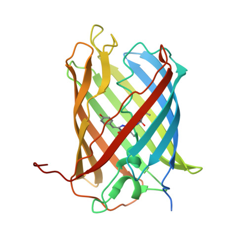
Orange Fluorescent Proteins: Structural Studies of LSSmOrange, PSmOrange and PSmOrange2.
Pletnev, S., Shcherbakova, D.M., Subach, O.M., Pletneva, N.V., Malashkevich, V.N., Almo, S.C., Dauter, Z., Verkhusha, V.V.(2014) PLoS One 9: e99136-e99136
- PubMed: 24960050
- DOI: https://doi.org/10.1371/journal.pone.0099136
- Primary Citation of Related Structures:
4Q7R, 4Q7T, 4Q7U - PubMed Abstract:
A structural analysis of the recently developed orange fluorescent proteins with novel phenotypes, LSSmOrange (λex/λem at 437/572 nm), PSmOrange (λex/λem at 548/565 nm and for photoconverted form at 636/662 nm) and PSmOrange2 (λex/λem at 546/561 nm and for photoconverted form at 619/651 nm), is presented. The obtained crystallographic structures provide an understanding of how the ensemble of a few key mutations enabled special properties of the orange FPs. While only a single Ile161Asp mutation, enabling excited state proton transfer, is critical for LSSmOrange, other substitutions provide refinement of its special properties and an exceptional 120 nm large Stokes shift. Similarly, a single Gln64Leu mutation was sufficient to cause structural changes resulting in photoswitchability of PSmOrange, and only one additional substitution (Phe65Ile), yielding PSmOrange2, was enough to greatly decrease the energy of photoconversion and increase its efficiency of photoswitching. Fluorescence of photoconverted PSmOrange and PSmOrange2 demonstrated an unexpected bathochromic shift relative to the fluorescence of classic red FPs, such as DsRed, eqFP578 and zFP574. The structural changes associated with this fluorescence shift are of considerable value for the design of advanced far-red FPs. For this reason the chromophore transformations accompanying photoconversion of the orange FPs are discussed.
Organizational Affiliation:
Leidos Biomedical Research Inc., Basic Research Program, Argonne, Illinois, United States of America; Macromolecular Crystallography Laboratory, National Cancer Institute, Argonne, Illinois, United States of America.