4Q2K

Bovine alpha chymotrypsin bound to a cyclic peptide inhibitor, 5b


Experimental Data Snapshot

  • Method: X-RAY DIFFRACTION
  • Resolution: 2.20 Å
  • R-Value Free: 0.268 
  • R-Value Work: 0.206 
  • R-Value Observed: 0.209 

wwPDB Validation   3D Report Full Report


Ligand Structure Quality Assessment 


This is version 1.2 of the entry. See complete history


Literature

Macrocyclic protease inhibitors with reduced peptide character.

Chua, K.C.Pietsch, M.Zhang, X.Hautmann, S.Chan, H.Y.Bruning, J.B.Gutschow, M.Abell, A.D.

(2014) Angew Chem Int Ed Engl 53: 7828-7831

  • DOI: https://doi.org/10.1002/anie.201404301
  • Primary Citation of Related Structures:  
    4Q2K

  • PubMed Abstract: 

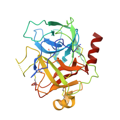
    There is a real need for simple structures that define a β-strand conformation, a secondary structure that is central to peptide-protein interactions. For example, protease substrates and inhibitors almost universally adopt this geometry on active site binding. A planar pyrrole is used to replace two amino acids of a peptide backbone to generate a simple macrocycle that retains the required geometry for active site binding. The resulting β-strand templates have reduced peptide character and provide potent protease inhibitors with the attachment of an appropriate amino aldehyde to the C-terminus. Picomolar inhibitors of cathepsin L and S are reported and the mode of binding of one example to the model protease chymotrypsin is defined by X-ray crystallography.


  • Organizational Affiliation

    School of Chemistry & Physics, The University of Adelaide, North Terrace, Adelaide, SA 5005 (Australia).


Macromolecules
Find similar proteins by:  (by identity cutoff)  |  3D Structure
Entity ID: 1
MoleculeChains Sequence LengthOrganismDetailsImage
Chymotrypsinogen A
A, B, C, D
245Bos taurusMutation(s): 0 
EC: 3.4.21.1
UniProt
Find proteins for P00766 (Bos taurus)
Explore P00766 
Go to UniProtKB:  P00766
Entity Groups  
Sequence Clusters30% Identity50% Identity70% Identity90% Identity95% Identity100% Identity
UniProt GroupP00766
Sequence Annotations
Expand
  • Reference Sequence
Small Molecules
Ligands 1 Unique
IDChains Name / Formula / InChI Key2D Diagram3D Interactions
5BF
Query on 5BF

Download Ideal Coordinates CCD File 
E [auth A],
F [auth B],
G [auth C],
H [auth D]
(11S)-4,9-dioxo-N-[(2S)-1-oxo-3-phenylpropan-2-yl]-17,22-dioxa-10,30-diazatetracyclo[21.2.2.2~13,16~.1~5,8~]triaconta-1(25),5,7,13,15,23,26,28-octaene-11-carboxamide
C36 H37 N3 O6
AGFHTNIIYCVXSL-UVMMSNCQSA-N
Experimental Data & Validation

Experimental Data

  • Method: X-RAY DIFFRACTION
  • Resolution: 2.20 Å
  • R-Value Free: 0.268 
  • R-Value Work: 0.206 
  • R-Value Observed: 0.209 
  • Space Group: P 1 21 1
Unit Cell:
Length ( Å )Angle ( ˚ )
a = 45.61α = 90
b = 109.34β = 93.47
c = 89.07γ = 90
Software Package:
Software NamePurpose
MOSFLMdata reduction
SCALAdata scaling
PHASERphasing
PHENIXrefinement
PDB_EXTRACTdata extraction

Structure Validation

View Full Validation Report


Ligand Structure Quality Assessment 


Entry History 

Deposition Data

Revision History  (Full details and data files)

  • Version 1.0: 2014-07-23
    Type: Initial release
  • Version 1.1: 2022-08-24
    Changes: Database references, Derived calculations
  • Version 1.2: 2023-11-08
    Changes: Data collection, Refinement description