4Q21

MOLECULAR SWITCH FOR SIGNAL TRANSDUCTION: STRUCTURAL DIFFERENCES BETWEEN ACTIVE AND INACTIVE FORMS OF PROTOONCOGENIC RAS PROTEINS


Experimental Data Snapshot

  • Method: X-RAY DIFFRACTION
  • Resolution: 2.00 Å
  • R-Value Work: 0.195 

wwPDB Validation   3D Report Full Report


This is version 1.4 of the entry. See complete history


Literature

Molecular switch for signal transduction: structural differences between active and inactive forms of protooncogenic ras proteins.

Milburn, M.V.Tong, L.deVos, A.M.Brunger, A.Yamaizumi, Z.Nishimura, S.Kim, S.H.

(1990) Science 247: 939-945

  • DOI: https://doi.org/10.1126/science.2406906
  • Primary Citation of Related Structures:  
    4Q21, 6Q21

  • PubMed Abstract: 

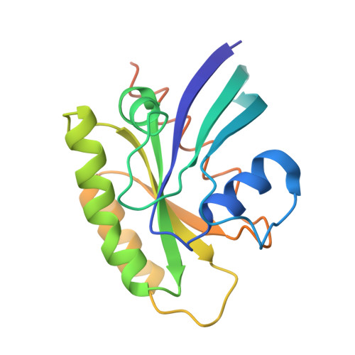
    Ras proteins participate as a molecular switch in the early steps of the signal transduction pathway that is associated with cell growth and differentiation. When the protein is in its GTP complexed form it is active in signal transduction, whereas it is inactive in its GDP complexed form. A comparison of eight three-dimensional structures of ras proteins in four different crystal lattices, five with a nonhydrolyzable GTP analog and three with GDP, reveals that the "on" and "off" states of the switch are distinguished by conformational differences that span a length of more than 40 A, and are induced by the gamma-phosphate. The most significant differences are localized in two regions: residues 30 to 38 (the switch I region) in the second loop and residues 60 to 76 (the switch II region) consisting of the fourth loop and the short alpha-helix that follows the loop. Both regions are highly exposed and form a continuous strip on the molecular surface most likely to be the recognition sites for the effector and receptor molecule(or molecules). The conformational differences also provide a structural basis for understanding the biological and biochemical changes of the proteins due to oncogenic mutations, autophosphorylation, and GTP hydrolysis, and for understanding the interactions with other proteins.


  • Organizational Affiliation

    Department of Chemistry, University of California, Berkeley.


Macromolecules
Find similar proteins by:  (by identity cutoff)  |  3D Structure
Entity ID: 1
MoleculeChains Sequence LengthOrganismDetailsImage
C-H-RAS P21 PROTEIN CATALYTIC DOMAIN189Homo sapiensMutation(s): 0 
UniProt & NIH Common Fund Data Resources
Find proteins for P01112 (Homo sapiens)
Explore P01112 
Go to UniProtKB:  P01112
PHAROS:  P01112
GTEx:  ENSG00000174775 
Entity Groups  
Sequence Clusters30% Identity50% Identity70% Identity90% Identity95% Identity100% Identity
UniProt GroupP01112
Sequence Annotations
Expand
  • Reference Sequence
Small Molecules
Ligands 2 Unique
IDChains Name / Formula / InChI Key2D Diagram3D Interactions
GDP
Query on GDP

Download Ideal Coordinates CCD File 
C [auth A]GUANOSINE-5'-DIPHOSPHATE
C10 H15 N5 O11 P2
QGWNDRXFNXRZMB-UUOKFMHZSA-N
MG
Query on MG

Download Ideal Coordinates CCD File 
B [auth A]MAGNESIUM ION
Mg
JLVVSXFLKOJNIY-UHFFFAOYSA-N
Experimental Data & Validation

Experimental Data

  • Method: X-RAY DIFFRACTION
  • Resolution: 2.00 Å
  • R-Value Work: 0.195 
  • Space Group: I 4
Unit Cell:
Length ( Å )Angle ( ˚ )
a = 96.3α = 90
b = 96.3β = 90
c = 41.37γ = 90
Software Package:
Software NamePurpose
X-PLORrefinement

Structure Validation

View Full Validation Report


Entry History 

Deposition Data

  • Released Date: 1992-07-15 
  • Deposition Author(s): Kim, S.-H.

Revision History  (Full details and data files)

  • Version 1.0: 1992-07-15
    Type: Initial release
  • Version 1.1: 2008-03-25
    Changes: Version format compliance
  • Version 1.2: 2011-07-13
    Changes: Version format compliance
  • Version 1.3: 2017-11-29
    Changes: Derived calculations, Other
  • Version 1.4: 2024-02-28
    Changes: Data collection, Database references, Derived calculations