4PY8

Crystal structure of Fab 3.1 in complex with the 1918 influenza virus hemagglutinin


Experimental Data Snapshot

  • Method: X-RAY DIFFRACTION
  • Resolution: 2.91 Å
  • R-Value Free: 0.242 
  • R-Value Work: 0.193 
  • R-Value Observed: 0.196 

wwPDB Validation   3D Report Full Report


Ligand Structure Quality Assessment 


This is version 2.0 of the entry. See complete history


Literature

Alternative Recognition of the Conserved Stem Epitope in Influenza A Virus Hemagglutinin by a VH3-30-Encoded Heterosubtypic Antibody.

Wyrzucki, A.Dreyfus, C.Kohler, I.Steck, M.Wilson, I.A.Hangartner, L.

(2014) J Virol 88: 7083-7092

  • DOI: https://doi.org/10.1128/JVI.00178-14
  • Primary Citation of Related Structures:  
    4PY7, 4PY8

  • PubMed Abstract: 

    A human monoclonal heterosubtypic antibody, MAb 3.1, with its heavy chain encoded by VH3-30, was isolated using phage display with immobilized hemagglutinin (HA) from influenza virus A/Japan/305/1957(H2N2) as the target. Antibody 3.1 potently neutralizes influenza viruses from the H1a clade (i.e., H1, H2, H5, H6) but has little neutralizing activity against the H1b clade. Its crystal structure in complex with HA from a pandemic H1N1 influenza virus, A/South Carolina/1/1918(H1N1), revealed that like other heterosubtypic anti-influenza virus antibodies, MAb 3.1 contacts a hydrophobic groove in the HA stem, primarily using its heavy chain. However, in contrast to the closely related monoclonal antibody (Mab) FI6 that relies heavily on HCDR3 for binding, MAb 3.1 utilizes residues from HCDR1, HCDR3, and framework region 3 (FR3). Interestingly, HCDR1 of MAb 3.1 adopts an α-helical conformation and engages in hydrophobic interactions with the HA very similar to those of the de novo in silico-designed and affinity-matured synthetic protein HB36.3. These findings improve our understanding of the molecular requirements for binding to the conserved epitope in the stem of the HA protein and, therefore, aid the development of more universal influenza vaccines targeting these epitopes.


  • Organizational Affiliation

    Institute of Medical Virology, University of Zurich, Zürich, Switzerland.


Macromolecules
Find similar proteins by:  (by identity cutoff)  |  3D Structure
Entity ID: 1
MoleculeChains Sequence LengthOrganismDetailsImage
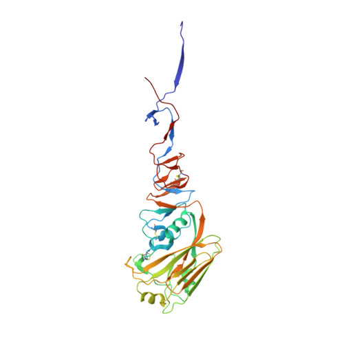
Hemagglutinin HA1 chain331Influenza A virus (A/South Carolina/1/1918(H1N1))Mutation(s): 0 
Gene Names: HAhemagglutinin
UniProt
Find proteins for Q9WFX3 (Influenza A virus (strain A/Brevig Mission/1/1918 H1N1))
Explore Q9WFX3 
Go to UniProtKB:  Q9WFX3
Entity Groups  
Sequence Clusters30% Identity50% Identity70% Identity90% Identity95% Identity100% Identity
UniProt GroupQ9WFX3
Sequence Annotations
Expand
  • Reference Sequence
Find similar proteins by:  (by identity cutoff)  |  3D Structure
Entity ID: 2
MoleculeChains Sequence LengthOrganismDetailsImage
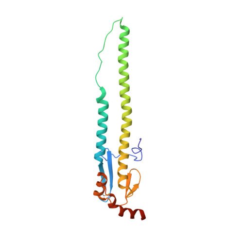
Hemagglutinin HA2 chain179Influenza A virus (A/South Carolina/1/1918(H1N1))Mutation(s): 0 
Gene Names: HAhemagglutinin
UniProt
Find proteins for Q9WFX3 (Influenza A virus (strain A/Brevig Mission/1/1918 H1N1))
Explore Q9WFX3 
Go to UniProtKB:  Q9WFX3
Entity Groups  
Sequence Clusters30% Identity50% Identity70% Identity90% Identity95% Identity100% Identity
UniProt GroupQ9WFX3
Sequence Annotations
Expand
  • Reference Sequence
Find similar proteins by:  (by identity cutoff)  |  3D Structure
Entity ID: 3
MoleculeChains Sequence LengthOrganismDetailsImage
antibody 3.1 heavy chainC [auth I]219Homo sapiensMutation(s): 0 
Entity Groups  
Sequence Clusters30% Identity50% Identity70% Identity90% Identity95% Identity100% Identity
Sequence Annotations
Expand
  • Reference Sequence
Find similar proteins by:  (by identity cutoff)  |  3D Structure
Entity ID: 4
MoleculeChains Sequence LengthOrganismDetailsImage
antibody 3.1 light chainD [auth J]214Homo sapiensMutation(s): 0 
Entity Groups  
Sequence Clusters30% Identity50% Identity70% Identity90% Identity95% Identity100% Identity
Sequence Annotations
Expand
  • Reference Sequence
Oligosaccharides

Help

Entity ID: 5
MoleculeChains Length2D Diagram Glycosylation3D Interactions
2-acetamido-2-deoxy-beta-D-glucopyranose-(1-4)-2-acetamido-2-deoxy-beta-D-glucopyranoseE [auth C]2N-Glycosylation
Glycosylation Resources
GlyTouCan:  G42666HT
GlyCosmos:  G42666HT
GlyGen:  G42666HT
Experimental Data & Validation

Experimental Data

  • Method: X-RAY DIFFRACTION
  • Resolution: 2.91 Å
  • R-Value Free: 0.242 
  • R-Value Work: 0.193 
  • R-Value Observed: 0.196 
  • Space Group: H 3
Unit Cell:
Length ( Å )Angle ( ˚ )
a = 135.06α = 90
b = 135.06β = 90
c = 230.201γ = 120
Software Package:
Software NamePurpose
Blu-Icedata collection
PHASERphasing
REFMACrefinement
HKL-2000data reduction
SCALEPACKdata scaling

Structure Validation

View Full Validation Report


Ligand Structure Quality Assessment 


Entry History 

Deposition Data

  • Released Date: 2014-05-21 
  • Deposition Author(s): Dreyfus, C.

Revision History  (Full details and data files)

  • Version 1.0: 2014-05-21
    Type: Initial release
  • Version 1.1: 2014-06-04
    Changes: Database references
  • Version 2.0: 2020-07-29
    Type: Remediation
    Reason: Carbohydrate remediation
    Changes: Advisory, Atomic model, Data collection, Database references, Derived calculations, Structure summary