4PQW

Crystal Structure of Phospholipase C beta 3 in Complex with PDZ1 of NHERF1


Experimental Data Snapshot

  • Method: X-RAY DIFFRACTION
  • Resolution: 1.47 Å
  • R-Value Free: 0.196 
  • R-Value Work: 0.179 
  • R-Value Observed: 0.180 

wwPDB Validation   3D Report Full Report


This is version 1.2 of the entry. See complete history


Literature

Crystallographic analysis of NHERF1-PLC beta 3 interaction provides structural basis for CXCR2 signaling in pancreatic cancer.

Jiang, Y.Wang, S.Holcomb, J.Trescott, L.Guan, X.Hou, Y.Brunzelle, J.Sirinupong, N.Li, C.Yang, Z.

(2014) Biochem Biophys Res Commun 446: 638-643

  • DOI: https://doi.org/10.1016/j.bbrc.2014.03.028
  • Primary Citation of Related Structures:  
    4PQW

  • PubMed Abstract: 

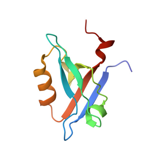
    The formation of CXCR2-NHERF1-PLCβ3 macromolecular complex in pancreatic cancer cells regulates CXCR2 signaling activity and plays an important role in tumor proliferation and invasion. We previously have shown that disruption of the NHERF1-mediated CXCR2-PLCβ3 interaction abolishes the CXCR2 signaling cascade and inhibits pancreatic tumor growth in vitro and in vivo. Here we report the crystal structure of the NHERF1 PDZ1 domain in complex with the C-terminal PLCβ3 sequence. The structure reveals that the PDZ1-PLCβ3 binding specificity is achieved by numerous hydrogen bonds and hydrophobic contacts with the last four PLCβ3 residues contributing to specific interactions. We also show that PLCβ3 can bind both NHERF1 PDZ1 and PDZ2 in pancreatic cancer cells, consistent with the observation that the peptide binding pockets of these PDZ domains are highly structurally conserved. This study provides an understanding of the structural basis for the PDZ-mediated NHERF1-PLCβ3 interaction that could prove valuable in selective drug design against CXCR2-related cancers.


  • Organizational Affiliation

    Department of Biochemistry and Molecular Biology, Wayne State University School of Medicine, Detroit, MI, USA.


Macromolecules
Find similar proteins by:  (by identity cutoff)  |  3D Structure
Entity ID: 1
MoleculeChains Sequence LengthOrganismDetailsImage
Na(+)/H(+) exchange regulatory cofactor NHE-RF191Homo sapiensMutation(s): 0 
Gene Names: NHERFNHERF1SLC9A3R1
UniProt & NIH Common Fund Data Resources
Find proteins for O14745 (Homo sapiens)
Explore O14745 
Go to UniProtKB:  O14745
PHAROS:  O14745
GTEx:  ENSG00000109062 
Entity Groups  
Sequence Clusters30% Identity50% Identity70% Identity90% Identity95% Identity100% Identity
UniProt GroupO14745
Sequence Annotations
Expand
  • Reference Sequence
Experimental Data & Validation

Experimental Data

  • Method: X-RAY DIFFRACTION
  • Resolution: 1.47 Å
  • R-Value Free: 0.196 
  • R-Value Work: 0.179 
  • R-Value Observed: 0.180 
  • Space Group: P 31 2 1
Unit Cell:
Length ( Å )Angle ( ˚ )
a = 50.66α = 90
b = 50.66β = 90
c = 66.68γ = 120
Software Package:
Software NamePurpose
HKL-2000data collection
PHASERphasing
PHENIXrefinement
HKL-2000data reduction
HKL-2000data scaling

Structure Validation

View Full Validation Report


Entry History 

Deposition Data

Revision History  (Full details and data files)

  • Version 1.0: 2014-04-02
    Type: Initial release
  • Version 1.1: 2014-04-23
    Changes: Database references
  • Version 1.2: 2023-09-20
    Changes: Data collection, Database references, Derived calculations, Refinement description