4PHT

ATPase GspE in complex with the cytoplasmic domain of GspL from the Vibrio vulnificus type II Secretion system


Experimental Data Snapshot

  • Method: X-RAY DIFFRACTION
  • Resolution: 2.83 Å
  • R-Value Free: 0.280 
  • R-Value Work: 0.248 
  • R-Value Observed: 0.249 

wwPDB Validation   3D Report Full Report


Ligand Structure Quality Assessment 


This is version 1.3 of the entry. See complete history


Literature

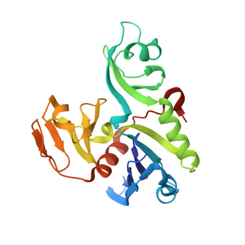
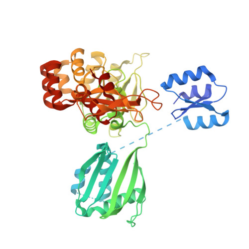
Crystal structure of the full-length ATPase GspE from the Vibrio vulnificus type II secretion system in complex with the cytoplasmic domain of GspL.

Lu, C.Korotkov, K.V.Hol, W.G.

(2014) J Struct Biol 187: 223-235

  • DOI: https://doi.org/10.1016/j.jsb.2014.07.006
  • Primary Citation of Related Structures:  
    4PHT

  • PubMed Abstract: 

    The type II secretion system (T2SS) is present in many Gram-negative bacteria and is responsible for secreting a large number of folded proteins, including major virulence factors, across the outer membrane. The T2SS consists of 11-15 different proteins most of which are present in multiple copies in the assembled secretion machinery. The ATPase GspE, essential for the functioning of the T2SS, contains three domains (N1E, N2E and CTE) of which the N1E domain is associated with the cytoplasmic domain of the inner membrane protein GspL. Here we describe and analyze the structure of the GspE•cyto-GspL complex from Vibrio vulnificus in the presence of an ATP analog, AMPPNP. There are three such ∼83 kDa complexes per asymmetric unit with essentially the same structure. The N2E and CTE domains of a single V. vulnificus GspE subunit adopt a mutual orientation that has not been seen before in any of the previous GspE structures, neither in structures of related ATPases from other secretion systems. This underlines the tremendous conformational flexibility of the T2SS secretion ATPase. Cyto-GspL interacts not only with the N1E domain, but also with the CTE domain and is even in contact with AMPPNP. Moreover, the cyto-GspL domains engage in two types of mutual interactions, resulting in two essentially identical, but crystallographically independent, "cyto-GspL rods" that run throughout the crystal. Very similar rods are present in previous crystals of cyto-GspL and of the N1E•cyto-GspL complex. This arrangement, now seen four times in three entirely different crystal forms, involves contacts between highly conserved residues suggesting a role in the biogenesis or the secretion mechanism or both of the T2SS.


  • Organizational Affiliation

    Department of Biochemistry and Biomolecular Structure Center, University of Washington, Seattle, WA 98195, United States.


Macromolecules
Find similar proteins by:  (by identity cutoff)  |  3D Structure
Entity ID: 1
MoleculeChains Sequence LengthOrganismDetailsImage
Type II secretion system protein LA [auth X],
C [auth Y],
E [auth Z]
246Vibrio vulnificus CMCP6Mutation(s): 0 
Gene Names: gspLVV1_0869
Entity Groups  
Sequence Clusters30% Identity50% Identity70% Identity90% Identity95% Identity100% Identity
Sequence Annotations
Expand
  • Reference Sequence
Find similar proteins by:  (by identity cutoff)  |  3D Structure
Entity ID: 2
MoleculeChains Sequence LengthOrganismDetailsImage
General secretory pathway protein EB [auth A],
D [auth B],
F [auth C]
500Vibrio vulnificus CMCP6Mutation(s): 0 
Gene Names: gspEVV1_0876
Entity Groups  
Sequence Clusters30% Identity50% Identity70% Identity90% Identity95% Identity100% Identity
Sequence Annotations
Expand
  • Reference Sequence
Small Molecules
Ligands 3 Unique
IDChains Name / Formula / InChI Key2D Diagram3D Interactions
ANP
Query on ANP

Download Ideal Coordinates CCD File 
H [auth A],
K [auth B],
N [auth C]
PHOSPHOAMINOPHOSPHONIC ACID-ADENYLATE ESTER
C10 H17 N6 O12 P3
PVKSNHVPLWYQGJ-KQYNXXCUSA-N
ZN
Query on ZN

Download Ideal Coordinates CCD File 
G [auth A],
J [auth B],
M [auth C]
ZINC ION
Zn
PTFCDOFLOPIGGS-UHFFFAOYSA-N
MG
Query on MG

Download Ideal Coordinates CCD File 
I [auth A],
L [auth B],
O [auth C]
MAGNESIUM ION
Mg
JLVVSXFLKOJNIY-UHFFFAOYSA-N
Experimental Data & Validation

Experimental Data

  • Method: X-RAY DIFFRACTION
  • Resolution: 2.83 Å
  • R-Value Free: 0.280 
  • R-Value Work: 0.248 
  • R-Value Observed: 0.249 
  • Space Group: C 1 2 1
Unit Cell:
Length ( Å )Angle ( ˚ )
a = 226.43α = 90
b = 133.9β = 91.41
c = 93.49γ = 90
Software Package:
Software NamePurpose
REFMACrefinement

Structure Validation

View Full Validation Report


Ligand Structure Quality Assessment 


Entry History & Funding Information

Deposition Data


Funding OrganizationLocationGrant Number
National Institutes of Health/National Institute of General Medical Sciences (NIH/NIGMS)United States--

Revision History  (Full details and data files)

  • Version 1.0: 2014-11-26
    Type: Initial release
  • Version 1.1: 2017-09-20
    Changes: Author supporting evidence, Derived calculations, Source and taxonomy
  • Version 1.2: 2019-12-25
    Changes: Author supporting evidence
  • Version 1.3: 2023-12-27
    Changes: Data collection, Database references, Derived calculations, Refinement description