4PFX

The highly conserved domain of unknown function 1792 has a distinct glycosyltransferase fold


Experimental Data Snapshot

  • Method: X-RAY DIFFRACTION
  • Resolution: 1.66 Å
  • R-Value Free: 0.192 
  • R-Value Work: 0.165 
  • R-Value Observed: 0.166 

wwPDB Validation   3D Report Full Report


Ligand Structure Quality Assessment 


This is version 1.5 of the entry. See complete history


Literature

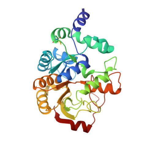
The highly conserved domain of unknown function 1792 has a distinct glycosyltransferase fold.

Zhang, H.Zhu, F.Yang, T.Ding, L.Zhou, M.Li, J.Haslam, S.M.Dell, A.Erlandsen, H.Wu, H.

(2014) Nat Commun 5: 4339-4339

  • DOI: https://doi.org/10.1038/ncomms5339
  • Primary Citation of Related Structures:  
    4PFX, 4PHR, 4PHS

  • PubMed Abstract: 

    More than 33,000 glycosyltransferases have been identified. Structural studies, however, have only revealed two distinct glycosyltransferase (GT) folds, GT-A and GT-B. Here we report a 1.34-Å resolution X-ray crystallographic structure of a previously uncharacterized 'domain of unknown function' 1792 (DUF1792) and show that the domain adopts a new fold and is required for glycosylation of a family of serine-rich repeat streptococcal adhesins. Biochemical studies reveal that the domain is a glucosyltransferase, and it catalyses the transfer of glucose to the branch point of the hexasaccharide O-linked to the serine-rich repeat of the bacterial adhesin, Fap1 of Streptococcus parasanguinis. DUF1792 homologues from both Gram-positive and Gram-negative bacteria also exhibit the activity. Thus, DUF1792 represents a new family of glycosyltransferases; therefore, we designate it as a GT-D glycosyltransferase fold. As the domain is highly conserved in bacteria and not found in eukaryotes, it can be explored as a new antibacterial target.


  • Organizational Affiliation

    Departments of Pediatric Dentistry, Microbiology, Schools of Dentistry and Medicine, University of Alabama at Birmingham, Birmingham, Alabama 35294, USA.


Macromolecules
Find similar proteins by:  (by identity cutoff)  |  3D Structure
Entity ID: 1
MoleculeChains Sequence LengthOrganismDetailsImage
Putative glycosyltransferase (GalT1)277Streptococcus parasanguinis FW213Mutation(s): 0 
Gene Names: galT1Spaf_1933
UniProt
Find proteins for I1ZPA1 (Streptococcus parasanguinis FW213)
Explore I1ZPA1 
Go to UniProtKB:  I1ZPA1
Entity Groups  
Sequence Clusters30% Identity50% Identity70% Identity90% Identity95% Identity100% Identity
UniProt GroupI1ZPA1
Sequence Annotations
Expand
  • Reference Sequence
Small Molecules
Ligands 2 Unique
IDChains Name / Formula / InChI Key2D Diagram3D Interactions
UDP
Query on UDP

Download Ideal Coordinates CCD File 
B [auth A]URIDINE-5'-DIPHOSPHATE
C9 H14 N2 O12 P2
XCCTYIAWTASOJW-XVFCMESISA-N
ACT
Query on ACT

Download Ideal Coordinates CCD File 
C [auth A]ACETATE ION
C2 H3 O2
QTBSBXVTEAMEQO-UHFFFAOYSA-M
Experimental Data & Validation

Experimental Data

  • Method: X-RAY DIFFRACTION
  • Resolution: 1.66 Å
  • R-Value Free: 0.192 
  • R-Value Work: 0.165 
  • R-Value Observed: 0.166 
  • Space Group: C 1 2 1
Unit Cell:
Length ( Å )Angle ( ˚ )
a = 71.337α = 90
b = 45.051β = 110.08
c = 78.915γ = 90
Software Package:
Software NamePurpose
PHENIXrefinement

Structure Validation

View Full Validation Report


Ligand Structure Quality Assessment 


Entry History & Funding Information

Deposition Data


Funding OrganizationLocationGrant Number
National Institutes of Health/National Institute of Dental and Craniofacial Research (NIH/NIDCR)United States5R01DE017954-07

Revision History  (Full details and data files)

  • Version 1.0: 2014-07-23
    Type: Initial release
  • Version 1.1: 2014-10-01
    Changes: Database references
  • Version 1.2: 2015-02-04
    Changes: Derived calculations
  • Version 1.3: 2017-09-27
    Changes: Author supporting evidence, Derived calculations, Other, Source and taxonomy
  • Version 1.4: 2019-12-11
    Changes: Author supporting evidence
  • Version 1.5: 2023-12-27
    Changes: Data collection, Database references, Refinement description