Functional and Structural Analysis of HicA3-HicB3, a Novel Toxin-Antitoxin System of Yersinia pestis.
Bibi-Triki, S., Li de la Sierra-Gallay, I., Lazar, N., Leroy, A., Van Tilbeurgh, H., Sebbane, F., Pradel, E.(2014) J Bacteriol 196: 3712-3723
- PubMed: 25112480
- DOI: https://doi.org/10.1128/JB.01932-14
- Primary Citation of Related Structures:
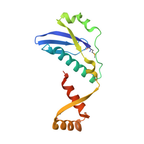
4P78, 4P7D - PubMed Abstract:
The mechanisms involved in the virulence of Yersinia pestis, the plague pathogen, are not fully understood. In previous research, we found that a Y. pestis mutant lacking the HicB3 (YPO3369) putative orphan antitoxin was attenuated for virulence in a murine model of bubonic plague. Toxin-antitoxin systems (TASs) are widespread in prokaryotes. Most bacterial species possess many TASs of several types. In type II TASs, the toxin protein is bound and neutralized by its cognate antitoxin protein in the cytoplasm. Here we identify the hicA3 gene encoding the toxin neutralized by HicB3 and show that HicA3-HicB3 constitutes a new functional type II TAS in Y. pestis. Using biochemical and mutagenesis-based approaches, we demonstrate that the HicA3 toxin is an RNase with a catalytic histidine residue. HicB3 has two functions: it sequesters and neutralizes HicA3 by blocking its active site, and it represses transcription of the hicA3B3 operon. Gel shift assays and reporter fusion experiments indicate that the HicB3 antitoxin binds to two operators in the hicA3B3 promoter region. We solved the X-ray structures of HicB3 and the HicA3-HicB3 complex; thus, we present the first crystal structure of a TA complex from the HicAB family. HicB3 forms a tetramer that can bind two HicA3 toxin molecules. HicA3 is monomeric and folds as a double-stranded-RNA-binding domain. The HicB3 N-terminal domain occludes the HicA3 active site, whereas its C-terminal domain folds as a ribbon-helix-helix DNA-binding motif.
Organizational Affiliation:
Equipe Peste et Yersinia pestis, INSERM U1019, Lille, France CNRS UMR 8204, Lille, France Institut Pasteur de Lille, Centre d'Infection et d'Immunité, Lille, France Université Lille Nord de France, Lille, France UDSL, Lille, France.