4P76

Cellular response to a crystal-forming protein


Experimental Data Snapshot

  • Method: X-RAY DIFFRACTION
  • Resolution: 2.90 Å
  • R-Value Free: 0.250 
  • R-Value Work: 0.174 
  • R-Value Observed: 0.178 

wwPDB Validation   3D Report Full Report


This is version 2.0 of the entry. See complete history


Literature

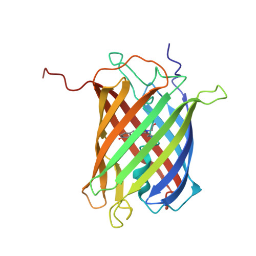
A diffraction-quality protein crystal processed as an autophagic cargo

Tsutsui, H.Jinno, Y.Shoda, K.Tomita, A.Matsuda, M.Yamashita, E.Katayama, H.Nakagawa, A.Miyawaki, A.

(2015) Mol Cell 58: 186-193

  • DOI: https://doi.org/10.1016/j.molcel.2015.02.007
  • Primary Citation of Related Structures:  
    4P76

  • PubMed Abstract: 

    Crystallization of proteins may occur in the cytosol of a living cell, but how a cell responds to intracellular protein crystallization remains unknown. We developed a variant of coral fluorescent protein that forms diffraction-quality crystals within mammalian cells. This expression system allowed the direct determination of its crystal structure at 2.9 Å, as well as observation of the crystallization process and cellular responses. The micron-sized crystal, which emerged rapidly, was a pure assembly of properly folded β-barrels and was recognized as an autophagic cargo that was transferred to lysosomes via a process involving p62 and LC3. Several lines of evidence indicated that autophagy was not required for crystal nucleation or growth. These findings demonstrate that in vivo protein crystals can provide an experimental model to study chemical catalysis. This knowledge may be beneficial for structural biology studies on normal and disease-related protein aggregation.


  • Organizational Affiliation

    Laboratory for Cell Function Dynamics, Brain Science Institute, RIKEN, Wako, Saitama 351-0198, Japan; Department of Material Science, Japan Advanced Institute of Science and Technology, Nomi, Ishikawa 923-1292, Japan; PRESTO, Japan Science and Technology Agency (JST), 4-1-8 Hon-cho, Kawaguchi, Saitama 351-0198, Japan.


Macromolecules
Find similar proteins by:  (by identity cutoff)  |  3D Structure
Entity ID: 1
MoleculeChains Sequence LengthOrganismDetailsImage
Photoconvertible fluorescent protein
A, B
226Dipsastraea favusMutation(s): 2 
Gene Names: KikGR
UniProt
Find proteins for Q53UG8 (Dipsastraea favus)
Explore Q53UG8 
Go to UniProtKB:  Q53UG8
Entity Groups  
Sequence Clusters30% Identity50% Identity70% Identity90% Identity95% Identity100% Identity
UniProt GroupQ53UG8
Sequence Annotations
Expand
  • Reference Sequence
Small Molecules
Ligands 1 Unique
IDChains Name / Formula / InChI Key2D Diagram3D Interactions
NA
Query on NA

Download Ideal Coordinates CCD File 
C [auth B]SODIUM ION
Na
FKNQFGJONOIPTF-UHFFFAOYSA-N
Modified Residues  1 Unique
IDChains TypeFormula2D DiagramParent
CR8
Query on CR8
A, B
L-PEPTIDE LINKINGC17 H16 N5 O4HIS, TYR, GLY
Experimental Data & Validation

Experimental Data

  • Method: X-RAY DIFFRACTION
  • Resolution: 2.90 Å
  • R-Value Free: 0.250 
  • R-Value Work: 0.174 
  • R-Value Observed: 0.178 
  • Space Group: P 21 21 2
Unit Cell:
Length ( Å )Angle ( ˚ )
a = 49.862α = 90
b = 84.426β = 90
c = 118.218γ = 90
Software Package:
Software NamePurpose
REFMACrefinement

Structure Validation

View Full Validation Report


Entry History 

Deposition Data

Revision History  (Full details and data files)

  • Version 1.0: 2015-04-29
    Type: Initial release
  • Version 1.1: 2020-01-22
    Changes: Data collection, Database references, Derived calculations, Source and taxonomy
  • Version 2.0: 2023-11-15
    Changes: Atomic model, Data collection, Database references, Derived calculations, Refinement description