


Crystal structure of the bacterial A1408C-mutant ribosomal decoding site
Kondo, J., Koganei, M.To be published.
Experimental Data Snapshot
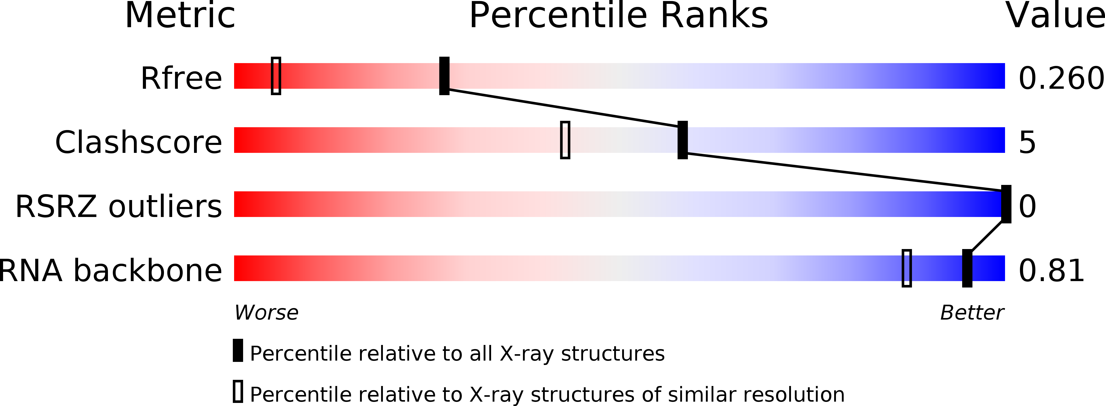
wwPDB Validation 3D Report Full Report
Find similar nucleic acids by: Sequence | 3D Structure
Entity ID: 1 | |||||
|---|---|---|---|---|---|
| Molecule | Chains | Length | Organism | Image | |
| 5'-D(*UP*UP*GP*CP*GP*UP*CP*CP*CP*GP*(5BU)P*CP*GP*AP*CP*GP*AP*AP*GP*UP*CP*GP*C)-3' | 23 | synthetic construct | 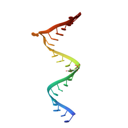 | ||
Sequence AnnotationsExpand | |||||
| |||||
| Length ( Å ) | Angle ( ˚ ) |
|---|---|
| a = 33.437 | α = 90 |
| b = 40.475 | β = 103.07 |
| c = 41.272 | γ = 90 |
| Software Name | Purpose |
|---|---|
| CrystalClear | data collection |
| PHASES | phasing |
| CNS | refinement |
| PDB_EXTRACT | data extraction |
| SOLVE | phasing |
| d*TREK | data reduction |
| d*TREK | data scaling |
| Funding Organization | Location | Grant Number |
|---|---|---|
| MEXT | Japan | 23790054 |
RCSB PDB (citation) is hosted by
RCSB PDB is a member of the

