4P2Q

Crystal structure of the 5cc7 TCR in complex with 5c2/I-Ek


Experimental Data Snapshot

  • Method: X-RAY DIFFRACTION
  • Resolution: 3.30 Å
  • R-Value Free: 0.236 
  • R-Value Work: 0.188 
  • R-Value Observed: 0.191 

wwPDB Validation   3D Report Full Report


This is version 1.8 of the entry. See complete history


Literature

Deconstructing the Peptide-MHC Specificity of T Cell Recognition.

Birnbaum, M.E.Mendoza, J.L.Sethi, D.K.Dong, S.Glanville, J.Dobbins, J.Ozkan, E.Davis, M.M.Wucherpfennig, K.W.Garcia, K.C.

(2014) Cell 157: 1073-1087

  • DOI: https://doi.org/10.1016/j.cell.2014.03.047
  • Primary Citation of Related Structures:  
    4P2O, 4P2Q, 4P2R

  • PubMed Abstract: 

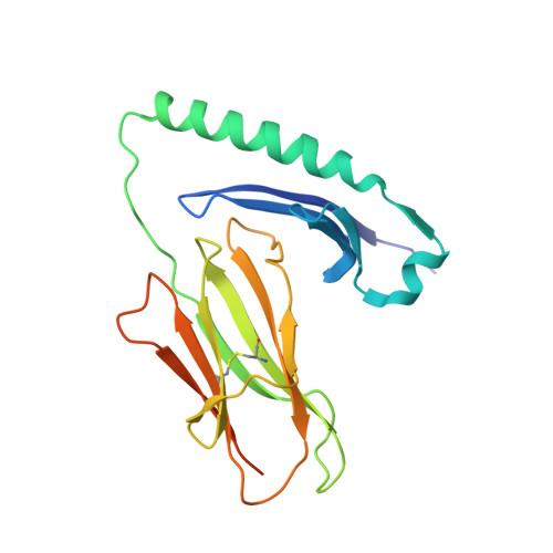
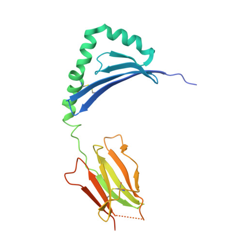
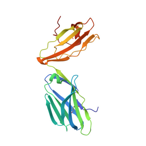
    In order to survey a universe of major histocompatibility complex (MHC)-presented peptide antigens whose numbers greatly exceed the diversity of the T cell repertoire, T cell receptors (TCRs) are thought to be cross-reactive. However, the nature and extent of TCR cross-reactivity has not been conclusively measured experimentally. We developed a system to identify MHC-presented peptide ligands by combining TCR selection of highly diverse yeast-displayed peptide-MHC libraries with deep sequencing. Although we identified hundreds of peptides reactive with each of five different mouse and human TCRs, the selected peptides possessed TCR recognition motifs that bore a close resemblance to their known antigens. This structural conservation of the TCR interaction surface allowed us to exploit deep-sequencing information to computationally identify activating microbial and self-ligands for human autoimmune TCRs. The mechanistic basis of TCR cross-reactivity described here enables effective surveillance of diverse self and foreign antigens without necessitating degenerate recognition of nonhomologous peptides.


  • Organizational Affiliation

    Departments of Molecular and Cellular Physiology and Structural Biology, Stanford University School of Medicine, Stanford, CA 94305, USA; Program in Immunology, Stanford University School of Medicine, Stanford, CA 94305, USA.


Macromolecules
Find similar proteins by:  (by identity cutoff)  |  3D Structure
Entity ID: 1
MoleculeChains Sequence LengthOrganismDetailsImage
H-2 class II histocompatibility antigen, E-K alpha chain
A, F, K, P
204Mus musculusMutation(s): 0 
UniProt
Find proteins for P04224 (Mus musculus)
Explore P04224 
Go to UniProtKB:  P04224
Entity Groups  
Sequence Clusters30% Identity50% Identity70% Identity90% Identity95% Identity100% Identity
UniProt GroupP04224
Sequence Annotations
Expand
  • Reference Sequence
Find similar proteins by:  (by identity cutoff)  |  3D Structure
Entity ID: 2
MoleculeChains Sequence LengthOrganismDetailsImage
MHC class II E-beta-k
B, G, L, Q
212Mus musculusMutation(s): 0 
Gene Names: H2-Eb1
UniProt & NIH Common Fund Data Resources
Find proteins for Q31163 (Mus musculus)
Explore Q31163 
Go to UniProtKB:  Q31163
IMPC:  MGI:95901
Entity Groups  
Sequence Clusters30% Identity50% Identity70% Identity90% Identity95% Identity100% Identity
UniProt GroupQ31163
Sequence Annotations
Expand
  • Reference Sequence

Find similar proteins by:  Sequence   |   3D Structure  

Entity ID: 3
MoleculeChains Sequence LengthOrganismDetailsImage
5c2 peptide
C, H, M, R
14synthetic constructMutation(s): 0 
Entity Groups  
Sequence Clusters30% Identity50% Identity70% Identity90% Identity95% Identity100% Identity
Sequence Annotations
Expand
  • Reference Sequence
Find similar proteins by:  (by identity cutoff)  |  3D Structure
Entity ID: 4
MoleculeChains Sequence LengthOrganismDetailsImage
5cc7 T-cell receptor alpha chain
D, I, N, S
205Mus musculusMutation(s): 0 
Entity Groups  
Sequence Clusters30% Identity50% Identity70% Identity90% Identity95% Identity100% Identity
Sequence Annotations
Expand
  • Reference Sequence
Find similar proteins by:  (by identity cutoff)  |  3D Structure
Entity ID: 5
MoleculeChains Sequence LengthOrganismDetailsImage
5cc7 T-cell receptor beta chain
E, J, O, T
266Mus musculusMutation(s): 0 
Entity Groups  
Sequence Clusters30% Identity50% Identity70% Identity90% Identity95% Identity100% Identity
Sequence Annotations
Expand
  • Reference Sequence
Experimental Data & Validation

Experimental Data

  • Method: X-RAY DIFFRACTION
  • Resolution: 3.30 Å
  • R-Value Free: 0.236 
  • R-Value Work: 0.188 
  • R-Value Observed: 0.191 
  • Space Group: C 1 2 1
Unit Cell:
Length ( Å )Angle ( ˚ )
a = 262.9α = 90
b = 102.21β = 95.04
c = 214.11γ = 90
Software Package:
Software NamePurpose
PHENIXrefinement

Structure Validation

View Full Validation Report


Entry History & Funding Information

Deposition Data


Funding OrganizationLocationGrant Number
National Institutes of Health/National Institute of General Medical Sciences (NIH/NIGMS)United States--

Revision History  (Full details and data files)

  • Version 1.0: 2014-05-21
    Type: Initial release
  • Version 1.1: 2014-06-11
    Changes: Database references
  • Version 1.2: 2015-01-14
    Changes: Database references
  • Version 1.3: 2015-02-25
    Changes: Derived calculations
  • Version 1.4: 2017-09-20
    Changes: Author supporting evidence, Derived calculations, Other, Source and taxonomy, Structure summary
  • Version 1.5: 2017-11-01
    Changes: Author supporting evidence
  • Version 1.6: 2019-12-25
    Changes: Author supporting evidence
  • Version 1.7: 2020-07-29
    Type: Remediation
    Reason: Carbohydrate remediation
    Changes: Data collection, Derived calculations, Refinement description, Structure summary
  • Version 1.8: 2023-12-27
    Changes: Data collection, Database references, Structure summary